 Movie
Movie Controller

[English] 日本語
 Yorodumi
- PDB-6qi5: Near Atomic Structure of an Atadenovirus Shows a possible gene du... -
+ Open data

- Basic information
| Entry | Database: PDB / ID: 6qi5 | ||||||
|---|---|---|---|---|---|---|---|
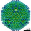
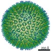
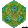
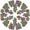
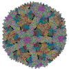
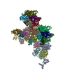
| Title | Near Atomic Structure of an Atadenovirus Shows a possible gene duplication event and Intergenera Variations in Cementing Proteins | ||||||
 Components Components |
| ||||||
 Keywords Keywords | VIRUS / adenovirus atadenovirus virus evolution | ||||||
| Function / homology |  Function and homology information Function and homology informationhexon binding / viral capsid, decoration / T=25 icosahedral viral capsid / microtubule-dependent intracellular transport of viral material towards nucleus / viral capsid / host cell / endocytosis involved in viral entry into host cell / symbiont entry into host cell / virion attachment to host cell / host cell nucleus / structural molecule activity Similarity search - Function | ||||||
| Biological species |  Lizard adenovirus 2 Lizard adenovirus 2 | ||||||
| Method | ELECTRON MICROSCOPY / single particle reconstruction / cryo EM / Resolution: 3.4 Å | ||||||
 Authors Authors | Condezo, G.N. / Marabini, R. / Gomez-Blanco, J. / SanMartin, C. | ||||||
 Citation Citation | Journal: Sci Adv / Year: 2021 Title: Near-atomic structure of an atadenovirus reveals a conserved capsid-binding motif and intergenera variations in cementing proteins. Authors: Roberto Marabini / Gabriela N Condezo / Mart Krupovic / Rosa Menéndez-Conejero / Josué Gómez-Blanco / Carmen San Martín /   Abstract: Of five known adenovirus genera, high-resolution structures are available only for mammalian-infecting mastadenoviruses. We present the first high-resolution structure of an adenovirus with ...Of five known adenovirus genera, high-resolution structures are available only for mammalian-infecting mastadenoviruses. We present the first high-resolution structure of an adenovirus with nonmammalian host: lizard atadenovirus LAdV-2. We find a large conformational difference in the internal vertex protein IIIa between mast- and atadenoviruses, induced by the presence of an extended polypeptide. This polypeptide, and α-helical clusters beneath the facet, likely correspond to genus-specific proteins LH2 and p32k. Another genus-specific protein, LH3, with a fold typical of bacteriophage tailspikes, contacts the capsid surface via a triskelion structure identical to that used by mastadenovirus protein IX, revealing a conserved capsid-binding motif and an ancient gene duplication event. Our data also suggest that mastadenovirus E1B-55 K was exapted from the atadenovirus-like LH3 protein. This work provides new information on the evolution of adenoviruses, emphasizing the importance of minor coat proteins for determining specific physicochemical properties of virions and most likely their tropism. #1: Journal: Biorxiv / Year: 2020Title: Near Atomic Structure of an Atadenovirus Reveals a Conserved Capsid-Binding Motif and Intergenera Variations in Cementing Proteins Authors: Marabini, R. / Condezo, G.N. / Gomez-Blanco, J. / San Martin, C. | ||||||
| History |
|
- Structure visualization
| Movie |
Movie viewer |
|---|---|
| Structure viewer | Molecule: MolmilJmol/JSmol |
- Downloads & links
-Download
| PDBx/mmCIF format | 6qi5.cif.gz | 2.3 MB | Display | PDBx/mmCIF format |
|---|---|---|---|---|
| PDB format | pdb6qi5.ent.gz | Display | PDB format | |
| PDBx/mmJSON format | 6qi5.json.gz | Tree view | PDBx/mmJSON format | |
| Others |  Other downloads Other downloads |
-Validation report
| Arichive directory | https://data.pdbj.org/pub/pdb/validation_reports/qi/6qi5ftp://data.pdbj.org/pub/pdb/validation_reports/qi/6qi5 | HTTPS FTP |
|---|
-Related structure data
| Related structure data |  4551MC M: map data used to model this data C: citing same article ( |
|---|---|
| Similar structure data |

-Links
 PDBj
PDBj


- Assembly
| Deposited unit | 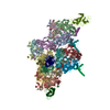
|
|---|---|
| 1 | x 60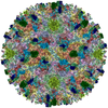
|
| Symmetry | Point symmetry: (Schoenflies symbol: I (icosahedral)) |
-Components
| #1: Protein | Mass: 101861.930 Da / Num. of mol.: 12 / Source method: isolated from a natural source / Source: (natural) Lizard adenovirus 2 / References: UniProt: A0A076FYV7#2: Protein | Mass: 41158.344 Da / Num. of mol.: 4 / Source method: isolated from a natural source / Source: (natural) Lizard adenovirus 2 / References: UniProt: A0A076FYU8#3: Protein | Mass: 30744.453 Da / Num. of mol.: 2 / Source method: isolated from a natural source / Source: (natural) Lizard adenovirus 2 / References: UniProt: A0A076FT36#4: Protein | | Mass: 67049.266 Da / Num. of mol.: 1 / Source method: isolated from a natural source / Source: (natural) Lizard adenovirus 2 / References: UniProt: A0A076FYV2#5: Protein | | Mass: 50619.848 Da / Num. of mol.: 1 / Source method: isolated from a natural source / Source: (natural) Lizard adenovirus 2 / References: UniProt: A0A076FT28 |
|---|
-Experimental details
-Experiment
| Experiment | Method: ELECTRON MICROSCOPY |
|---|---|
| EM experiment | Aggregation state: PARTICLE / 3D reconstruction method: single particle reconstruction |
- Sample preparation
| Component | Name: Lizard adenovirus 2 / Type: VIRUS / Entity ID: all / Source: NATURAL |
|---|---|
| Molecular weight | Value: 150 MDa / Experimental value: NO |
| Source (natural) | Organism: Lizard adenovirus 2 |
| Details of virus | Empty: NO / Enveloped: NO / Isolate: SPECIES / Type: VIRION |
| Natural host | Organism: Heloderma horridum |
| Virus shell | Diameter: 940 nm / Triangulation number (T number): 25 |
| Buffer solution | pH: 7.4 / Details: PBS |
| Specimen | Embedding applied: NO / Shadowing applied: NO / Staining applied: NO / Vitrification applied: YES |
| Specimen support | Grid material: COPPER/RHODIUM / Grid type: Quantifoil R2/4 |
| Vitrification | Cryogen name: ETHANE |
- Electron microscopy imaging
| Experimental equipment |  Model: Titan Krios / Image courtesy: FEI Company |
|---|---|
| Microscopy | Model: FEI TITAN KRIOS |
| Electron gun | Electron source:  FIELD EMISSION GUN / Accelerating voltage: 300 kV / Illumination mode: FLOOD BEAM FIELD EMISSION GUN / Accelerating voltage: 300 kV / Illumination mode: FLOOD BEAM |
| Electron lens | Mode: BRIGHT FIELD |
| Image recording | Average exposure time: 2 sec. / Electron dose: 54 e/Å2 / Detector mode: INTEGRATING / Film or detector model: FEI FALCON II (4k x 4k) |
- Processing
| Software |
| ||||||||||||||||||||||||||||||||||||||||||||||||||||||
|---|---|---|---|---|---|---|---|---|---|---|---|---|---|---|---|---|---|---|---|---|---|---|---|---|---|---|---|---|---|---|---|---|---|---|---|---|---|---|---|---|---|---|---|---|---|---|---|---|---|---|---|---|---|---|---|
| EM software |
| ||||||||||||||||||||||||||||||||||||||||||||||||||||||
| CTF correction | Type: PHASE FLIPPING ONLY | ||||||||||||||||||||||||||||||||||||||||||||||||||||||
| Symmetry | Point symmetry: I (icosahedral) | ||||||||||||||||||||||||||||||||||||||||||||||||||||||
| 3D reconstruction | Resolution: 3.4 Å / Resolution method: FSC 0.143 CUT-OFF / Num. of particles: 16071 / Symmetry type: POINT | ||||||||||||||||||||||||||||||||||||||||||||||||||||||
| Refinement | Stereochemistry target values: GeoStd + Monomer Library | ||||||||||||||||||||||||||||||||||||||||||||||||||||||
| Refine LS restraints |
|
