 Movie
Movie Controller

[English] 日本語
 Yorodumi
- EMDB-4551: Near Atomic Structure of an Atadenovirus Shows a possible gene du... -
+ Open data

- Basic information
| Entry | Database: EMDB / ID: EMD-4551 | |||||||||
|---|---|---|---|---|---|---|---|---|---|---|
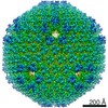
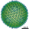
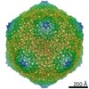
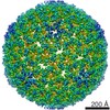
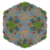
| Title | Near Atomic Structure of an Atadenovirus Shows a possible gene duplication event and Intergenera Variations in Cementing Proteins | |||||||||
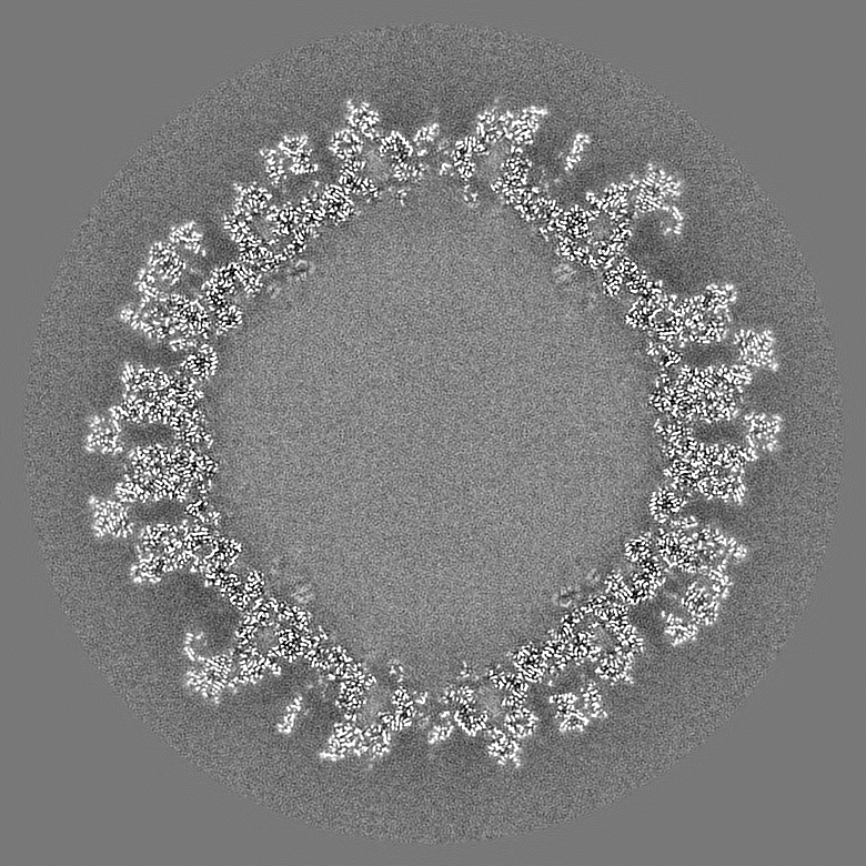
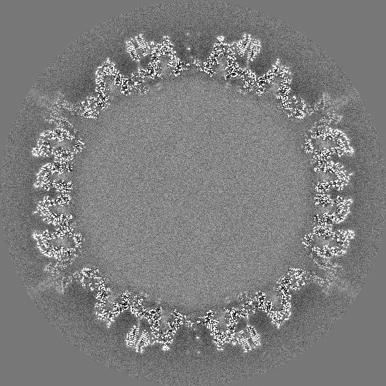
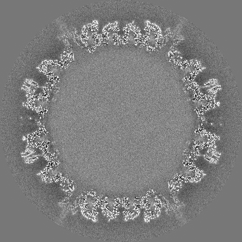
 Map data Map data | Cryo-EM structure of a lizard atadenovirus, LAdV-2, at 3.4 A resolution | |||||||||
 Sample Sample |
| |||||||||
 Keywords Keywords | adenovirus atadenovirus virus evolution / VIRUS | |||||||||
| Function / homology |  Function and homology information Function and homology informationhexon binding / viral capsid, decoration / T=25 icosahedral viral capsid / microtubule-dependent intracellular transport of viral material towards nucleus / viral capsid / host cell / endocytosis involved in viral entry into host cell / symbiont entry into host cell / virion attachment to host cell / host cell nucleus / structural molecule activity Similarity search - Function | |||||||||
| Biological species |  Lizard adenovirus 2 Lizard adenovirus 2 | |||||||||
| Method | single particle reconstruction / cryo EM / Resolution: 3.4 Å | |||||||||
 Authors Authors | Condezo GN / Marabini R | |||||||||
 Citation Citation | Journal: Sci Adv / Year: 2021 Title: Near-atomic structure of an atadenovirus reveals a conserved capsid-binding motif and intergenera variations in cementing proteins. Authors: Roberto Marabini / Gabriela N Condezo / Mart Krupovic / Rosa Menéndez-Conejero / Josué Gómez-Blanco / Carmen San Martín /   Abstract: Of five known adenovirus genera, high-resolution structures are available only for mammalian-infecting mastadenoviruses. We present the first high-resolution structure of an adenovirus with ...Of five known adenovirus genera, high-resolution structures are available only for mammalian-infecting mastadenoviruses. We present the first high-resolution structure of an adenovirus with nonmammalian host: lizard atadenovirus LAdV-2. We find a large conformational difference in the internal vertex protein IIIa between mast- and atadenoviruses, induced by the presence of an extended polypeptide. This polypeptide, and α-helical clusters beneath the facet, likely correspond to genus-specific proteins LH2 and p32k. Another genus-specific protein, LH3, with a fold typical of bacteriophage tailspikes, contacts the capsid surface via a triskelion structure identical to that used by mastadenovirus protein IX, revealing a conserved capsid-binding motif and an ancient gene duplication event. Our data also suggest that mastadenovirus E1B-55 K was exapted from the atadenovirus-like LH3 protein. This work provides new information on the evolution of adenoviruses, emphasizing the importance of minor coat proteins for determining specific physicochemical properties of virions and most likely their tropism. #1: Journal: Biorxiv / Year: 2020Title: Near Atomic Structure of an Atadenovirus Reveals a Conserved Capsid-Binding Motif and Intergenera Variations in Cementing Proteins Authors: Marabini R / Condezo GN / Gomez-Blanco J / San Martin C | |||||||||
| History |
|
- Structure visualization
| Movie |
Movie viewer |
|---|---|
| Structure viewer | EM map: SurfViewMolmilJmol/JSmol |
| Supplemental images |

- Downloads & links
-EMDB archive
| Map data | emd_4551.map.gz | 1.6 GB | EMDB map data format | |
|---|---|---|---|---|
| Header (meta data) | emd-4551-v30.xmlemd-4551.xml | 17.1 KB 17.1 KB | Display Display | EMDB header |
| FSC (resolution estimation) | emd_4551_fsc.xml | 29.6 KB | Display | FSC data file |
| Images | 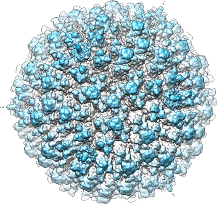 emd_4551.png emd_4551.png | 314.4 KB | ||
| Filedesc metadata | emd-4551.cif.gz | 7.3 KB | ||
| Archive directory |  http://ftp.pdbj.org/pub/emdb/structures/EMD-4551ftp://ftp.pdbj.org/pub/emdb/structures/EMD-4551 http://ftp.pdbj.org/pub/emdb/structures/EMD-4551ftp://ftp.pdbj.org/pub/emdb/structures/EMD-4551 | HTTPS FTP |
-Related structure data
| Related structure data |  6qi5MC M: atomic model generated by this map C: citing same article ( |
|---|---|
| Similar structure data |


-Links
| EMDB pages | EMDB (EBI/PDBe) / EMDataResource |
|---|---|
| Related items in Molecule of the Month |


-Map
| File | Download / File: emd_4551.map.gz / Format: CCP4 / Size: 1.8 GB / Type: IMAGE STORED AS FLOATING POINT NUMBER (4 BYTES) | ||||||||||||||||||||||||||||||||||||||||||||||||||||||||||||||||||||
|---|---|---|---|---|---|---|---|---|---|---|---|---|---|---|---|---|---|---|---|---|---|---|---|---|---|---|---|---|---|---|---|---|---|---|---|---|---|---|---|---|---|---|---|---|---|---|---|---|---|---|---|---|---|---|---|---|---|---|---|---|---|---|---|---|---|---|---|---|---|
| Annotation | Cryo-EM structure of a lizard atadenovirus, LAdV-2, at 3.4 A resolution | ||||||||||||||||||||||||||||||||||||||||||||||||||||||||||||||||||||
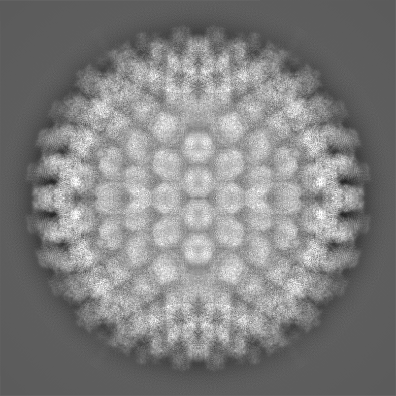
| Projections & slices | Image control
Images are generated by Spider. | ||||||||||||||||||||||||||||||||||||||||||||||||||||||||||||||||||||
| Voxel size | X=Y=Z: 1.35 Å | ||||||||||||||||||||||||||||||||||||||||||||||||||||||||||||||||||||
| Density |
| ||||||||||||||||||||||||||||||||||||||||||||||||||||||||||||||||||||
| Symmetry | Space group: 1 | ||||||||||||||||||||||||||||||||||||||||||||||||||||||||||||||||||||
| Details | EMDB XML:
CCP4 map header:
| ||||||||||||||||||||||||||||||||||||||||||||||||||||||||||||||||||||
 Z (Sec.)
 Y (Row.)
 X (Col.)


-Supplemental data
- Sample components
-Entire : Lizard adenovirus 2
| Entire | Name: Lizard adenovirus 2 |
|---|---|
| Components |
|
-Supramolecule #1: Lizard adenovirus 2
| Supramolecule | Name: Lizard adenovirus 2 / type: virus / ID: 1 / Parent: 0 / Macromolecule list: all / NCBI-ID: 874272 / Sci species name: Lizard adenovirus 2 / Virus type: VIRION / Virus isolate: SPECIES / Virus enveloped: No / Virus empty: No |
|---|---|
| Host (natural) | Organism:  Heloderma horridum (Mexican beaded lizard) Heloderma horridum (Mexican beaded lizard) |
| Molecular weight | Theoretical: 150 MDa |
| Virus shell | Shell ID: 1 / Diameter: 940.0 Å / T number (triangulation number): 25 |
-Macromolecule #1: Hexon protein
| Macromolecule | Name: Hexon protein / type: protein_or_peptide / ID: 1 / Number of copies: 12 / Enantiomer: LEVO |
|---|---|
| Source (natural) | Organism: Lizard adenovirus 2 |
| Molecular weight | Theoretical: 101.86193 KDa |
| Sequence | String: MEPQREFFHI AGRSAKEYLS ENLVQFIQAT QNYFNIGEKF RDPYVAPSAG VTTDRSQKLQ LRVVPIQTED NVNYYKARFT LNVGDNRLV DLGSSYFDIK GTLDRGPSFK PYGGTAYNPL APKSAPINSA FTVGNDTHFV AQLPQTYAAG GTGVTEAIQQ Q VSGVDPNP ...String: MEPQREFFHI AGRSAKEYLS ENLVQFIQAT QNYFNIGEKF RDPYVAPSAG VTTDRSQKLQ LRVVPIQTED NVNYYKARFT LNVGDNRLV DLGSSYFDIK GTLDRGPSFK PYGGTAYNPL APKSAPINSA FTVGNDTHFV AQLPQTYAAG GTGVTEAIQQ Q VSGVDPNP QVGQPNYAGP VVVNTTNNAG LGRIVSADSE GQQFPCYGAY APPQSAGGDV STAAVTKTYI NTTNNNGRVS GT MATDTIT WENPDAHFAD FVDDRRATAA GNRPNYIGFR DNFIGMMYYN SGSNTGSFSS QTQQLNIVLD LNDRNSELSY QYL LADLTS RWHYFALWNQ AVDDYDHHVR ILENDGYEEG PPNLAFPPHV ISNPFAPAAV GTGMTVNEQQ QTAAVTANTV ALIG YGNIP AVEMNLPANL KRTFLYSNVA MYLPDTYKFT PANVDLPENH LSYGYINGRL PLPNIVDTWT DIGARWSLDV MDTVN PFNH HRNTGLKYRS QLLGNGRYCD FHIQVPQKFF AIKNLLLLPG TYNYEWYFRK DPNMVLQSTL GNDLRADGAS ITYTQI NLY VSFFPMNYDT QSELELMLRN ATNDQNFSDY LGAVNNLYQI PAGSSTVVVN IPDRSWGAFR GWSFTRLKVS ETPRIGA TQ DPNFQYSGSI PYLDGTFYLS HTFQRCSIQW DSSVPWPGND RMLTPNWFEI KRPINQDAEG NDTMQSNLTK DFFMVQMA A SYNQGYQGFN WPNCTKHYGF INNFEPMSRQ VPEYGANYPN LMAAYLANPQ TMPIWNNCGF QQKTATNVLL ERCGHPYVA NWPYPLSGRN AVPNQVTERK FLVDRYLWQI PFSSNFLNMG TLTDLGQNVM YANSSHSLNM QFTVDPMTEP TYLMLLFGVF DQVVINQPT RSGISVAYLR LPFASGSAAT UniProtKB: Hexon protein |
-Macromolecule #2: Protein LH3
| Macromolecule | Name: Protein LH3 / type: protein_or_peptide / ID: 2 / Number of copies: 4 / Enantiomer: LEVO |
|---|---|
| Source (natural) | Organism: Lizard adenovirus 2 |
| Molecular weight | Theoretical: 41.158344 KDa |
| Sequence | String: MTSVEELYVI NPINQWPAPG SFSSQKPPGT LLPGEDPEAV FKQYHVVYLV PGAQYHWKNI LIEKPVWIYG NGATVRTSGT GPILRIVGN RTEKRDVRIQ DISFFGEDCT PNRMEPMSEK LVYQMAIWVT DMKRVTIKGC NFTNFAGAAV FFEETAYNGF F WSMQHLIT ...String: MTSVEELYVI NPINQWPAPG SFSSQKPPGT LLPGEDPEAV FKQYHVVYLV PGAQYHWKNI LIEKPVWIYG NGATVRTSGT GPILRIVGN RTEKRDVRIQ DISFFGEDCT PNRMEPMSEK LVYQMAIWVT DMKRVTIKGC NFTNFAGAAV FFEETAYNGF F WSMQHLIT ECRFTGCRIG IANGGRSEYS TASFNNFFDC QICFNVVGGN WNRCGNIAAN CRCVYLHTTN MWYEGAGGNF NA AHGSFTG NTMNHCDYGG NLWPTAFQLP DREIQLAGFY FDNARARCPT WTGNTQYYGD MKILNFNQAN DAAIFVIDGC ALY GQPGDT GSIETTAALT DKVFIQGCQG NKVTLFNIKA ANVVPAIGTI KQKP UniProtKB: Protein LH3 |
-Macromolecule #3: Pre-hexon-linking protein VIII
| Macromolecule | Name: Pre-hexon-linking protein VIII / type: protein_or_peptide / ID: 3 / Number of copies: 2 / Enantiomer: LEVO |
|---|---|
| Source (natural) | Organism: Lizard adenovirus 2 |
| Molecular weight | Theoretical: 30.744453 KDa |
| Sequence | String: MDAPVTPYIW QYQPQTGKAA GARQNYGAVI NWLSADNNMF HRVQTVNRAR NLIDEIREET VRPDLAASFN DWTYDQLTQP PGTAYLPAP DPLTGPTTIR DKVLSAEGEQ LAGSRPSVLH GGASLPPSAY SLGDGREYMK LTRDALPFPQ NWMVKENGVW T PIVEQRAA ...String: MDAPVTPYIW QYQPQTGKAA GARQNYGAVI NWLSADNNMF HRVQTVNRAR NLIDEIREET VRPDLAASFN DWTYDQLTQP PGTAYLPAP DPLTGPTTIR DKVLSAEGEQ LAGSRPSVLH GGASLPPSAY SLGDGREYMK LTRDALPFPQ NWMVKENGVW T PIVEQRAA LRGGSANALS SYPTLLFNQP PILRYRRPGQ QLQGSGVIAP SSKVLSLLSE APRIPRTEGM TPYQFANSFP PV VYEDPFS QNLAVFPKEF SPLFEPENQV LASSLATLQY N UniProtKB: Pre-hexon-linking protein VIII |
-Macromolecule #4: PIIIa
| Macromolecule | Name: PIIIa / type: protein_or_peptide / ID: 4 / Number of copies: 1 / Enantiomer: LEVO |
|---|---|
| Source (natural) | Organism: Lizard adenovirus 2 |
| Molecular weight | Theoretical: 67.049266 KDa |
| Sequence | String: MDPGLKPSSL WTHKIIDSII ANRSLSAVQN FRKQPLANKL TALEDAIVQP RKDTTPETVA AILQELVAMG ALQPNEVGPM FSDLMIRVH KYNSTNVQNN LSVLLGDIRA AQSEAIRSTN VGELSNQVVL NDFLSREPAV VPQGQHNYEA FKQTLRLMVN E APNVTLFK ...String: MDPGLKPSSL WTHKIIDSII ANRSLSAVQN FRKQPLANKL TALEDAIVQP RKDTTPETVA AILQELVAMG ALQPNEVGPM FSDLMIRVH KYNSTNVQNN LSVLLGDIRA AQSEAIRSTN VGELSNQVVL NDFLSREPAV VPQGQHNYEA FKQTLRLMVN E APNVTLFK SGPDTLMQVN IRGVNTVNLN SAFKNLKNFW GVQLDTEIVP GSISSKLSSN TRVLLLFLAP FTNDSTFTPD TF ISQIMRL YRETVAASIE QPQETELEVA ETIRELGGDV EDIGRTMAFL LKNKEEIVSN PRTLSPRQLN VLRYVQESLQ DRI DRNGEE PEDALRNLVF SFAPSYFEAN GPFIRRLISY LEVALINSPN YFREIYSNKY WTPPASFWTQ NYGDFHLERE AEAE RRAAS EAGYGDFEEG DFALPDNLGE GSDLAWDDFN TAMSPSVPPT PSVRSAPASL SYGRTSPSSV SSLTASDRNI GATLA RAVI PPAAAAIGSA AGEALYPSLG QYLAPAASLA ATRLLNLTRA RRQRLKRDSL ARHRRITEVR GVYPKAAPTR SSSSSS VSS TPTVFEPLPG AFSVINPLMR PEGDRDVSGT GIVNPFSHLK PRNGLQ UniProtKB: PIIIa |
-Macromolecule #5: Penton protein
| Macromolecule | Name: Penton protein / type: protein_or_peptide / ID: 5 / Number of copies: 1 / Enantiomer: LEVO |
|---|---|
| Source (natural) | Organism: Lizard adenovirus 2 |
| Molecular weight | Theoretical: 50.619848 KDa |
| Sequence | String: MEVYVPPPRV MAPTEGRNSI SYNPIAPLQD TTHIYIIDNK TSDIENLNIH KDHSNFYTNI VQNVDVAPSD AATQTIKLDE RSRWGGELH TILKTNAPNV TEFFNSNSFK ALLMSDKTDP ANPVYTWFEL SIPEGDYTVG SLIDMLNNAV VENYLEVGRQ K GVQISDIG ...String: MEVYVPPPRV MAPTEGRNSI SYNPIAPLQD TTHIYIIDNK TSDIENLNIH KDHSNFYTNI VQNVDVAPSD AATQTIKLDE RSRWGGELH TILKTNAPNV TEFFNSNSFK ALLMSDKTDP ANPVYTWFEL SIPEGDYTVG SLIDMLNNAV VENYLEVGRQ K GVQISDIG VKFDTRNFSL GRDPLTSLVT PGNYTFKAFH PDIVLLPGCG VDFTHSRINN MLGMRKRFPY EPGYVITYED LV GGNIPAL LDLAKYPGET SPVLQDPDGN SYHVEEVSPK KWQTKYRSWC LAYNSSQGTL KSEQILTVPD ITGGLGQLYW SLP DAFKPP VTFTNNTTDI STQPVTGMHL FPLSQRIVYN TSAVYAQLVE QMTNNTKVFN RFPKNAILMQ PPYDTTQWIS ENVP YVADH GIQPLKNSLT GVQRVTLTDD RRRSCPYIYK TLATVTPKVL SSATLQ UniProtKB: Penton protein |
-Experimental details
-Structure determination
| Method | cryo EM |
|---|---|
 Processing Processing | single particle reconstruction |
| Aggregation state | particle |
-Sample preparation
| Buffer | pH: 7.4 / Details: PBS |
|---|---|
| Grid | Model: Quantifoil R2/4 / Material: COPPER/RHODIUM |
| Vitrification | Cryogen name: ETHANE |
- Electron microscopy
| Microscope | FEI TITAN KRIOS |
|---|---|
| Image recording | Film or detector model: FEI FALCON II (4k x 4k) / Detector mode: INTEGRATING / Average exposure time: 2.0 sec. / Average electron dose: 54.0 e/Å2 |
| Electron beam | Acceleration voltage: 300 kV / Electron source:  FIELD EMISSION GUN FIELD EMISSION GUN |
| Electron optics | Illumination mode: FLOOD BEAM / Imaging mode: BRIGHT FIELD |
| Experimental equipment |  Model: Titan Krios / Image courtesy: FEI Company |

