 Movie
Movie Controller

[English] 日本語
 Yorodumi
- EMDB-5375: Direct electron detection yields cryo-EM reconstructions at resol... -
+ Open data

- Basic information
| Entry | Database: EMDB / ID: EMD-5375 | |||||||||
|---|---|---|---|---|---|---|---|---|---|---|
| Title | Direct electron detection yields cryo-EM reconstructions at resolutions beyond .75 Nyquist frequency | |||||||||
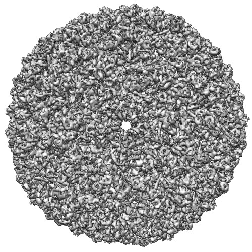
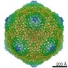
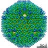
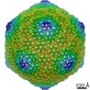
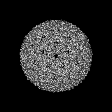
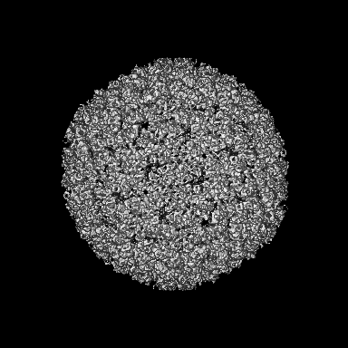
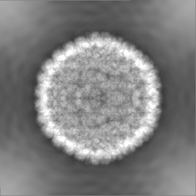
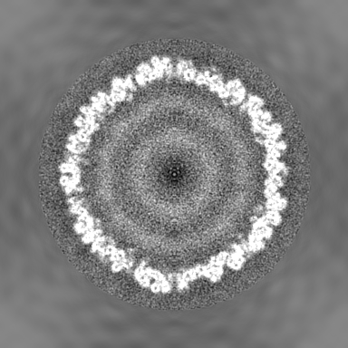
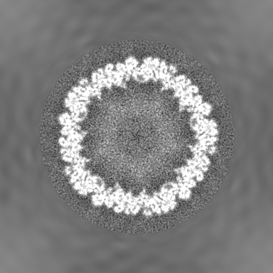
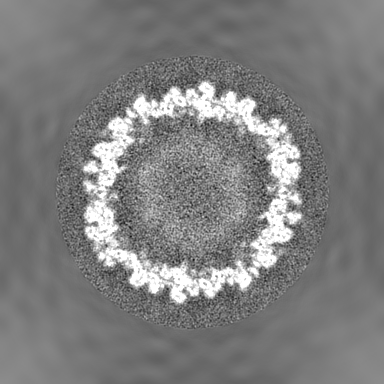
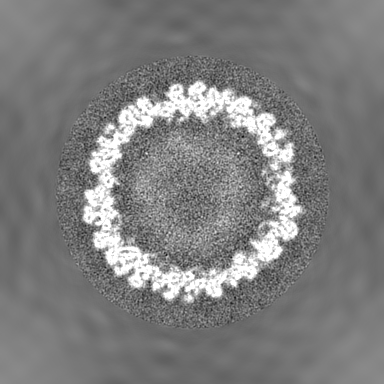
 Map data Map data | Icosahedral reconstruction of bacteriophage P22 | |||||||||
 Sample Sample |
| |||||||||
 Keywords Keywords | Cryo-EM / Electron cryo-microscopy / Direct detection device / Active pixel sensor / CMOS detector / Nyquist frequency | |||||||||
| Biological species |  Enterobacteria phage P22 (virus) Enterobacteria phage P22 (virus) | |||||||||
| Method | single particle reconstruction / cryo EM / Resolution: 8.5 Å | |||||||||
 Authors Authors | Bammes BE / Rochat RH / Jakana J / Chen D / Chiu W | |||||||||
 Citation Citation | Journal: J Struct Biol / Year: 2012 Title: Direct electron detection yields cryo-EM reconstructions at resolutions beyond 3/4 Nyquist frequency. Authors: Benjamin E Bammes / Ryan H Rochat / Joanita Jakana / Dong-Hua Chen / Wah Chiu /  Abstract: One limitation in electron cryo-microscopy (cryo-EM) is the inability to recover high-resolution signal from the image-recording media at the full-resolution limit of the transmission electron ...One limitation in electron cryo-microscopy (cryo-EM) is the inability to recover high-resolution signal from the image-recording media at the full-resolution limit of the transmission electron microscope. Direct electron detection using CMOS-based sensors for digitally recording images has the potential to alleviate this shortcoming. Here, we report a practical performance evaluation of a Direct Detection Device (DDD®) for biological cryo-EM at two different microscope voltages: 200 and 300 kV. Our DDD images of amorphous and graphitized carbon show strong per-pixel contrast with image resolution near the theoretical sampling limit of the data. Single-particle reconstructions of two frozen-hydrated bacteriophages, P22 and ε15, establish that the DDD is capable of recording usable signal for 3D reconstructions at about 4/5 of the Nyquist frequency, which is a vast improvement over the performance of conventional imaging media. We anticipate the unparalleled performance of this digital recording device will dramatically benefit cryo-EM for routine tomographic and single-particle structural determination of biological specimens. | |||||||||
| History |
|
- Structure visualization
| Movie |
 Movie viewer Movie viewer |
|---|---|
| Structure viewer | EM map: SurfViewMolmilJmol/JSmol |
| Supplemental images |
- Downloads & links
-EMDB archive
| Map data | emd_5375.map.gz | 196.4 MB | EMDB map data format | |
|---|---|---|---|---|
| Header (meta data) | emd-5375-v30.xmlemd-5375.xml | 9.9 KB 9.9 KB | Display Display | EMDB header |
| Images |  emd_5375_1.jpg emd_5375_1.jpg | 116.4 KB | ||
| Archive directory |  http://ftp.pdbj.org/pub/emdb/structures/EMD-5375ftp://ftp.pdbj.org/pub/emdb/structures/EMD-5375 http://ftp.pdbj.org/pub/emdb/structures/EMD-5375ftp://ftp.pdbj.org/pub/emdb/structures/EMD-5375 | HTTPS FTP |
-Related structure data


-Links
| EMDB pages | EMDB (EBI/PDBe) / EMDataResource |
|---|
-Map
| File | Download / File: emd_5375.map.gz / Format: CCP4 / Size: 210.9 MB / Type: IMAGE STORED AS FLOATING POINT NUMBER (4 BYTES) | ||||||||||||||||||||||||||||||||||||||||||||||||||||||||||||||||||||
|---|---|---|---|---|---|---|---|---|---|---|---|---|---|---|---|---|---|---|---|---|---|---|---|---|---|---|---|---|---|---|---|---|---|---|---|---|---|---|---|---|---|---|---|---|---|---|---|---|---|---|---|---|---|---|---|---|---|---|---|---|---|---|---|---|---|---|---|---|---|
| Annotation | Icosahedral reconstruction of bacteriophage P22 | ||||||||||||||||||||||||||||||||||||||||||||||||||||||||||||||||||||
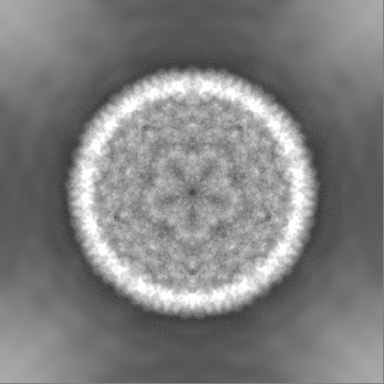
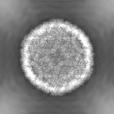
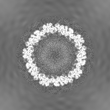
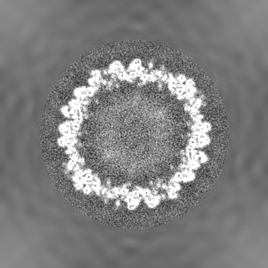
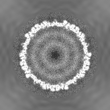
| Projections & slices | Image control
Images are generated by Spider. | ||||||||||||||||||||||||||||||||||||||||||||||||||||||||||||||||||||
| Voxel size | X=Y=Z: 3.48 Å | ||||||||||||||||||||||||||||||||||||||||||||||||||||||||||||||||||||
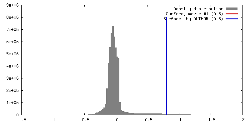
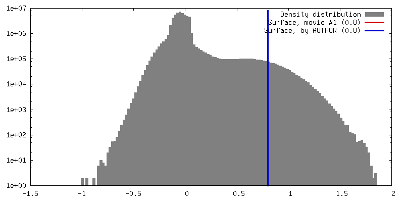
| Density |
| ||||||||||||||||||||||||||||||||||||||||||||||||||||||||||||||||||||
| Symmetry | Space group: 1 | ||||||||||||||||||||||||||||||||||||||||||||||||||||||||||||||||||||
| Details | EMDB XML:
CCP4 map header:
| ||||||||||||||||||||||||||||||||||||||||||||||||||||||||||||||||||||
 Z (Sec.)
 Y (Row.)
 X (Col.)


-Supplemental data
- Sample components
-Entire : Bacteriophage P22 procapsid
| Entire | Name: Bacteriophage P22 procapsid |
|---|---|
| Components |
|
-Supramolecule #1000: Bacteriophage P22 procapsid
| Supramolecule | Name: Bacteriophage P22 procapsid / type: sample / ID: 1000 / Number unique components: 1 |
|---|
-Supramolecule #1: Enterobacteria phage P22
| Supramolecule | Name: Enterobacteria phage P22 / type: virus / ID: 1 / Name.synonym: P22 / NCBI-ID: 10754 / Sci species name: Enterobacteria phage P22 / Database: NCBI / Virus type: VIRION / Virus isolate: STRAIN / Virus enveloped: No / Virus empty: Yes / Syn species name: P22 |
|---|---|
| Host (natural) | Organism:  Salmonella (bacteria) / synonym: BACTERIA(EUBACTERIA) Salmonella (bacteria) / synonym: BACTERIA(EUBACTERIA) |
| Virus shell | Shell ID: 1 / Name: Procapsid / Diameter: 525 Å / T number (triangulation number): 7 |
-Experimental details
-Structure determination
| Method | cryo EM |
|---|---|
 Processing Processing | single particle reconstruction |
| Aggregation state | particle |
-Sample preparation
| Concentration | 1 mg/mL |
|---|---|
| Buffer | pH: 7.6 / Details: 50 mM Tris pH 7.6, 25 mM NaCl, 2mM EDTA |
| Grid | Details: 400 mesh copper grid |
| Vitrification | Cryogen name: ETHANE / Chamber humidity: 95 % / Chamber temperature: 170 K / Instrument: OTHER / Details: Vitrification instrument: Vitrobot / Method: Blot for 2 seconds before plunging |
- Electron microscopy
| Microscope | JEOL 2010F |
|---|---|
| Temperature | Min: 100 K / Max: 100 K / Average: 100 K |
| Alignment procedure | Legacy - Astigmatism: objective lens astigmatism was corrected at 100,000 times magnification |
| Date | Jun 10, 2010 |
| Image recording | Category: CCD / Film or detector model: DIRECT ELECTRON DE-12 (4k x 3k) / Digitization - Sampling interval: 6 µm / Number real images: 200 / Average electron dose: 17 e/Å2 Details: Images were collected on a CMOS type detector DE-12 from Direct Electron |
| Electron beam | Acceleration voltage: 200 kV / Electron source:  FIELD EMISSION GUN FIELD EMISSION GUN |
| Electron optics | Calibrated magnification: 17200 / Illumination mode: FLOOD BEAM / Imaging mode: BRIGHT FIELD / Cs: 2.0 mm / Nominal defocus max: 5.0 µm / Nominal defocus min: 2.0 µm / Nominal magnification: 15000 |
| Sample stage | Specimen holder: Side Entry / Specimen holder model: GATAN LIQUID NITROGEN |
-Image processing
| CTF correction | Details: Each Micrograph |
|---|---|
| Final reconstruction | Algorithm: OTHER / Resolution.type: BY AUTHOR / Resolution: 8.5 Å / Resolution method: FSC 0.5 CUT-OFF / Software - Name: MPSA Details: Resolution was calculated to be 8.5 Angstroms at 0.5 FSC and 7.3 Angstroms at 0.143 FSC as compared against the 3.8 Angstrom reconstruction of P22. Number images used: 7500 |
