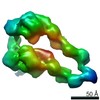
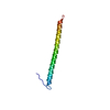
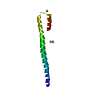
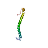
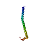
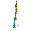
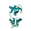
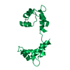
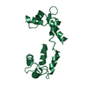
[日本語] English
- PDB-7qio: Homology model of myosin neck domain in skeletal sarcomere -

+
データを開く


IDまたはキーワード:

読み込み中...

-
基本情報

登録情報
データベース: PDB / ID: 7qio
タイトルHomology model of myosin neck domain in skeletal sarcomere
要素
  • Myosin light chain 1/3, skeletal muscle isoform
  • Myosin regulatory light chain 2, skeletal muscle isoform
  • Myosin-4
キーワードMOTOR PROTEIN / Skeletal muscle / Sarcomere / Cross-bridge
機能・相同性
機能・相同性情報


Striated Muscle Contraction / Smooth Muscle Contraction / contractile muscle fiber / myosin filament / myosin complex / structural constituent of muscle / myosin II complex / microfilament motor activity / myofibril / cardiac muscle contraction ...Striated Muscle Contraction / Smooth Muscle Contraction / contractile muscle fiber / myosin filament / myosin complex / structural constituent of muscle / myosin II complex / microfilament motor activity / myofibril / cardiac muscle contraction / skeletal muscle tissue development / response to activity / muscle contraction / double-stranded RNA binding / actin filament binding / calmodulin binding / immune response / calcium ion binding / ATP binding / cytoplasm
類似検索 - 分子機能
EF-hand domain / DNA repair protein XRCC4-like, C-terminal / Myosin tail / Myosin tail / Myosin N-terminal SH3-like domain / Myosin S1 fragment, N-terminal / Myosin, N-terminal, SH3-like / Myosin N-terminal SH3-like domain profile. / Myosin head, motor domain / Myosin head (motor domain) ...EF-hand domain / DNA repair protein XRCC4-like, C-terminal / Myosin tail / Myosin tail / Myosin N-terminal SH3-like domain / Myosin S1 fragment, N-terminal / Myosin, N-terminal, SH3-like / Myosin N-terminal SH3-like domain profile. / Myosin head, motor domain / Myosin head (motor domain) / Myosin motor domain profile. / Myosin. Large ATPases. / IQ motif profile. / IQ motif, EF-hand binding site / Kinesin motor domain superfamily / EF-hand, calcium binding motif / EF-Hand 1, calcium-binding site / EF-hand calcium-binding domain. / EF-hand calcium-binding domain profile. / EF-hand domain / EF-hand domain pair / P-loop containing nucleoside triphosphate hydrolase
類似検索 - ドメイン・相同性
Myosin light chain 1/3, skeletal muscle isoform / Myosin regulatory light chain 11 / Myosin-4
類似検索 - 構成要素
生物種Mus musculus (ハツカネズミ)
手法電子顕微鏡法 / サブトモグラム平均法 / クライオ電子顕微鏡法 / 解像度: 9 Å
データ登録者Wang, Z. / Grange, M. / Pospich, S. / Wagner, T. / Kho, A.L. / Gautel, M. / Raunser, S.
資金援助European Union, 5件
組織認可番号
Max Planck SocietyEuropean Union
Wellcome Trust201543/Z/16/ZEuropean Union
European Research Council (ERC)856118European Union
Medical Research Council (MRC, United Kingdom)MR/R003106/1European Union
European Molecular Biology Organization (EMBO)European Union
引用ジャーナル: Science / : 2022
タイトル: Structures from intact myofibrils reveal mechanism of thin filament regulation through nebulin.
著者: Zhexin Wang / Michael Grange / Sabrina Pospich / Thorsten Wagner / Ay Lin Kho / Mathias Gautel / Stefan Raunser /
要旨: In skeletal muscle, nebulin stabilizes and regulates the length of thin filaments, but the underlying mechanism remains nebulous. In this work, we used cryo-electron tomography and subtomogram ...In skeletal muscle, nebulin stabilizes and regulates the length of thin filaments, but the underlying mechanism remains nebulous. In this work, we used cryo-electron tomography and subtomogram averaging to reveal structures of native nebulin bound to thin filaments within intact sarcomeres. This in situ reconstruction provided high-resolution details of the interaction between nebulin and actin, demonstrating the stabilizing role of nebulin. Myosin bound to the thin filaments exhibited different conformations of the neck domain, highlighting its inherent structural variability in muscle. Unexpectedly, nebulin did not interact with myosin or tropomyosin, but it did interact with a troponin T linker through two potential binding motifs on nebulin, explaining its regulatory role. Our structures support the role of nebulin as a thin filament "molecular ruler" and provide a molecular basis for studying nemaline myopathies.
履歴
登録2021年12月15日登録サイト: PDBE / 処理サイト: PDBE
改定 1.02022年2月16日Provider: repository / タイプ: Initial release
改定 1.12022年3月2日Group: Database references / カテゴリ: citation
Item: _citation.journal_volume / _citation.page_first ..._citation.journal_volume / _citation.page_first / _citation.page_last / _citation.pdbx_database_id_PubMed / _citation.title
改定 1.22024年7月17日Group: Data collection / Refinement description
カテゴリ: chem_comp_atom / chem_comp_bond ...chem_comp_atom / chem_comp_bond / em_3d_fitting_list / em_admin / pdbx_initial_refinement_model
Item: _em_3d_fitting_list.accession_code / _em_3d_fitting_list.initial_refinement_model_id ..._em_3d_fitting_list.accession_code / _em_3d_fitting_list.initial_refinement_model_id / _em_3d_fitting_list.source_name / _em_3d_fitting_list.type / _em_admin.last_update

-
構造の表示

ムービー
  • 登録構造単位
  • Jmolによる作画
  • ダウンロード
  • EMマップとの重ね合わせ
  • マップデータ: EMDB-13993
  • UCSF Chimeraによる作画
  • ダウンロード
ムービービューア
構造ビューア分子:
MolmilJmol/JSmol

ダウンロードとリンク

-
集合体

登録構造単位
L: Myosin-4
M: Myosin-4
N: Myosin light chain 1/3, skeletal muscle isoform
O: Myosin light chain 1/3, skeletal muscle isoform
P: Myosin regulatory light chain 2, skeletal muscle isoform
Q: Myosin regulatory light chain 2, skeletal muscle isoform


分子量 (理論値)分子数
合計 (水以外)272,0096
ポリマ-272,0096
非ポリマー00
00
1
L: Myosin-4


分子量 (理論値)分子数
合計 (水以外)96,4061
ポリマ-96,4061
非ポリマー00
0
タイプ名称対称操作
identity operation1_5551
2
M: Myosin-4


分子量 (理論値)分子数
合計 (水以外)96,4061
ポリマ-96,4061
非ポリマー00
0
タイプ名称対称操作
identity operation1_5551
3
N: Myosin light chain 1/3, skeletal muscle isoform


分子量 (理論値)分子数
合計 (水以外)20,6201
ポリマ-20,6201
非ポリマー00
0
タイプ名称対称操作
identity operation1_5551
4
O: Myosin light chain 1/3, skeletal muscle isoform


分子量 (理論値)分子数
合計 (水以外)20,6201
ポリマ-20,6201
非ポリマー00
0
タイプ名称対称操作
identity operation1_5551
5
P: Myosin regulatory light chain 2, skeletal muscle isoform


分子量 (理論値)分子数
合計 (水以外)18,9781
ポリマ-18,9781
非ポリマー00
0
タイプ名称対称操作
identity operation1_5551
6
Q: Myosin regulatory light chain 2, skeletal muscle isoform


分子量 (理論値)分子数
合計 (水以外)18,9781
ポリマ-18,9781
非ポリマー00
0
タイプ名称対称操作
identity operation1_5551

-
要素

#1: タンパク質 Myosin-4 / Myosin heavy chain 2b / MyHC-2b / Myosin heavy chain 4


分子量: 96405.609 Da / 分子数: 2 / 由来タイプ: 天然 / 由来: (天然) Mus musculus (ハツカネズミ) / 参照: UniProt: Q5SX39
#2: タンパク質 Myosin light chain 1/3, skeletal muscle isoform / MLC1/MLC3 / MLC1F/MLC3F / Myosin light chain alkali 1/2 / Myosin light chain A1/A2


分子量: 20620.490 Da / 分子数: 2 / 由来タイプ: 天然 / 由来: (天然) Mus musculus (ハツカネズミ) / 参照: UniProt: P05977
#3: タンパク質 Myosin regulatory light chain 2, skeletal muscle isoform / Fast skeletal myosin light chain 2 / MLC2F


分子量: 18978.445 Da / 分子数: 2 / 由来タイプ: 天然 / 由来: (天然) Mus musculus (ハツカネズミ) / 参照: UniProt: P97457

-
実験情報

-
実験

実験手法: 電子顕微鏡法
EM実験試料の集合状態: TISSUE / 3次元再構成法: サブトモグラム平均法

-
試料調製

構成要素名称: Mouse psoas muscle myofibrils / タイプ: ORGANELLE OR CELLULAR COMPONENT / Entity ID: all / 由来: NATURAL
由来(天然)生物種: Mus musculus (ハツカネズミ)
緩衝液pH: 7
試料包埋: NO / シャドウイング: NO / 染色: NO / 凍結: YES
急速凍結装置: FEI VITROBOT MARK IV / 凍結剤: ETHANE

-
電子顕微鏡撮影

実験機器
モデル: Titan Krios / 画像提供: FEI Company
顕微鏡モデル: FEI TITAN KRIOS
電子銃電子線源: FIELD EMISSION GUN / 加速電圧: 300 kV / 照射モード: FLOOD BEAM
電子レンズモード: BRIGHT FIELD / 倍率(公称値): 81000 X / 最大 デフォーカス(公称値): 5000 nm / 最小 デフォーカス(公称値): 2400 nm / Calibrated defocus min: 2400 nm / 最大 デフォーカス(補正後): 5000 nm / Cs: 2.7 mm / C2レンズ絞り径: 50 µm
試料ホルダ凍結剤: NITROGEN
試料ホルダーモデル: FEI TITAN KRIOS AUTOGRID HOLDER
撮影電子線照射量: 3.4 e/Å2 / Avg electron dose per subtomogram: 130 e/Å2 / 検出モード: COUNTING
フィルム・検出器のモデル: GATAN K2 SUMMIT (4k x 4k)

-
解析

EMソフトウェア
ID名称カテゴリ
2SerialEM画像取得
7UCSF Chimeraモデルフィッティング
CTF補正タイプ: PHASE FLIPPING AND AMPLITUDE CORRECTION
対称性点対称性: C1 (非対称)
3次元再構成解像度: 9 Å / 解像度の算出法: FSC 0.143 CUT-OFF / 粒子像の数: 44001 / 対称性のタイプ: POINT
EM volume selectionNum. of tomograms: 48 / Num. of volumes extracted: 183260
原子モデル構築B value: 300 / プロトコル: RIGID BODY FIT / 空間: REAL
原子モデル構築

3D fitting-ID: 1 / Source name: PDB / タイプ: experimental model

IDPDB-IDPDB chain-IDAccession codeInitial refinement model-ID
17QINL7QIN1
23I5GB3I5G2
33I5GC3I5G2

+
万見について

-
お知らせ

-
2022年2月9日: EMDBエントリの付随情報ファイルのフォーマットが新しくなりました

EMDBエントリの付随情報ファイルのフォーマットが新しくなりました

  • EMDBのヘッダファイルのバージョン3が、公式のフォーマットとなりました。
  • これまでは公式だったバージョン1.9は、アーカイブから削除されます。

関連情報:EMDBヘッダ

外部リンク:wwPDBはEMDBデータモデルのバージョン3へ移行します

-
2020年8月12日: 新型コロナ情報

新型コロナ情報

URL: https://pdbjlvh1.pdbj.org/emnavi/covid19.php

新ページ: EM Navigatorに新型コロナウイルスの特設ページを開設しました。

関連情報:Covid-19情報 / 2020年3月5日: 新型コロナウイルスの構造データ

+
2020年3月5日: 新型コロナウイルスの構造データ

新型コロナウイルスの構造データ

関連情報:万見生物種 / 2020年8月12日: 新型コロナ情報

外部リンク:COVID-19特集ページ - PDBj / 今月の分子2020年2月:コロナウイルスプロテーアーゼ

+
2019年1月31日: EMDBのIDの桁数の変更

EMDBのIDの桁数の変更

  • EMDBエントリに付与されているアクセスコード(EMDB-ID)は4桁の数字(例、EMD-1234)でしたが、間もなく枯渇します。これまでの4桁のID番号は4桁のまま変更されませんが、4桁の数字を使い切った後に発行されるIDは5桁以上の数字(例、EMD-12345)になります。5桁のIDは2019年の春頃から発行される見通しです。
  • EM Navigator/万見では、接頭語「EMD-」は省略されています。

関連情報:Q: 「EMD」とは何ですか? / 万見/EM NavigatorにおけるID/アクセスコードの表記

外部リンク:EMDB Accession Codes are Changing Soon! / PDBjへお問い合わせ

+
2017年7月12日: PDB大規模アップデート

PDB大規模アップデート

  • 新バージョンのPDBx/mmCIF辞書形式に基づくデータがリリースされました。
  • 今回の更新はバージョン番号が4から5になる大規模なもので、全エントリデータの書き換えが行われる「Remediation」というアップデートに該当します。
  • このバージョンアップで、電子顕微鏡の実験手法に関する多くの項目の書式が改定されました(例:em_softwareなど)。
  • EM NavigatorとYorodumiでも、この改定に基づいた表示内容になります。

外部リンク:wwPDB Remediation / OneDepデータ基準に準拠した、より強化された内容のモデル構造ファイルが、PDBアーカイブで公開されました。

-
万見 (Yorodumi)

幾万の構造データを、幾万の視点から

  • 万見(Yorodumi)は、EMDB/PDB/SASBDBなどの構造データを閲覧するためのページです。
  • EM Navigatorの詳細ページの後継、Omokage検索のフロントエンドも兼ねています。

関連情報:EMDB / PDB / SASBDB / 3つのデータバンクの比較 / 万見検索 / 2016年8月31日: 新しいEM Navigatorと万見 / 万見文献 / Jmol/JSmol / 機能・相同性情報 / 新しいEM Navigatorと万見の変更点

他の情報も見る