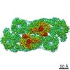
Journal: Science / Year: 2022 Title: Structures from intact myofibrils reveal mechanism of thin filament regulation through nebulin. Authors: Zhexin Wang / Michael Grange / Sabrina Pospich / Thorsten Wagner / Ay Lin Kho / Mathias Gautel / Stefan Raunser / Abstract: In skeletal muscle, nebulin stabilizes and regulates the length of thin filaments, but the underlying mechanism remains nebulous. In this work, we used cryo-electron tomography and subtomogram ...In skeletal muscle, nebulin stabilizes and regulates the length of thin filaments, but the underlying mechanism remains nebulous. In this work, we used cryo-electron tomography and subtomogram averaging to reveal structures of native nebulin bound to thin filaments within intact sarcomeres. This in situ reconstruction provided high-resolution details of the interaction between nebulin and actin, demonstrating the stabilizing role of nebulin. Myosin bound to the thin filaments exhibited different conformations of the neck domain, highlighting its inherent structural variability in muscle. Unexpectedly, nebulin did not interact with myosin or tropomyosin, but it did interact with a troponin T linker through two potential binding motifs on nebulin, explaining its regulatory role. Our structures support the role of nebulin as a thin filament "molecular ruler" and provide a molecular basis for studying nemaline myopathies.
History
Deposition
Dec 15, 2021
-
Header (metadata) release
Feb 16, 2022
-
Map release
Feb 16, 2022
-
Update
Mar 4, 2026
-
Current status
Mar 4, 2026
Processing site: PDBe / Status: Released
-
Structure visualization
Movie
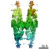
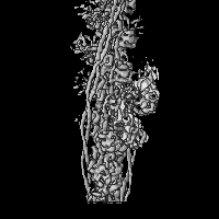
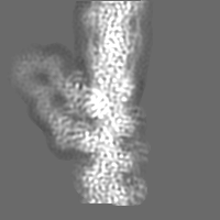
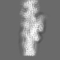
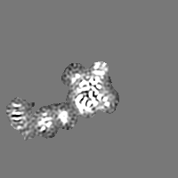
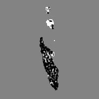
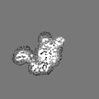
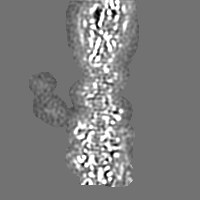
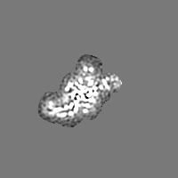
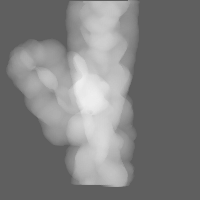
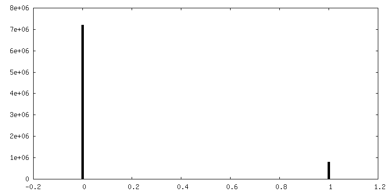
Surface view with section colored by density value
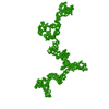
Name: nebulin (mouse) / type: protein_or_peptide / ID: 2 Details: Model with conserved tyrosines of three nebulin simple repeats Number of copies: 1 / Enantiomer: LEVO
Name: nebulin (mouse) / type: protein_or_peptide / ID: 3 Details: Model with conserved tyrosines of two nebulin simple repeats Number of copies: 1 / Enantiomer: LEVO
In the structure databanks used in Yorodumi, some data are registered as the other names, "COVID-19 virus" and "2019-nCoV". Here are the details of the virus and the list of structure data.
Jan 31, 2019. EMDB accession codes are about to change! (news from PDBe EMDB page)
EMDB accession codes are about to change! (news from PDBe EMDB page)
The allocation of 4 digits for EMDB accession codes will soon come to an end. Whilst these codes will remain in use, new EMDB accession codes will include an additional digit and will expand incrementally as the available range of codes is exhausted. The current 4-digit format prefixed with “EMD-” (i.e. EMD-XXXX) will advance to a 5-digit format (i.e. EMD-XXXXX), and so on. It is currently estimated that the 4-digit codes will be depleted around Spring 2019, at which point the 5-digit format will come into force.
The EM Navigator/Yorodumi systems omit the EMD- prefix.
Related info.:Q: What is EMD? / ID/Accession-code notation in Yorodumi/EM Navigator
Yorodumi is a browser for structure data from EMDB, PDB, SASBDB, etc.
This page is also the successor to EM Navigator detail page, and also detail information page/front-end page for Omokage search.
The word "yorodu" (or yorozu) is an old Japanese word meaning "ten thousand". "mi" (miru) is to see.
Related info.:EMDB / PDB / SASBDB / Comparison of 3 databanks / Yorodumi Search / Aug 31, 2016. New EM Navigator & Yorodumi / Yorodumi Papers / Jmol/JSmol / Function and homology information / Changes in new EM Navigator and Yorodumi
 Movie
Movie Controller


 Open data
 Basic information
 Map data
 Sample
 Keywords
 Function and homology information
 Authors
 Citation

 Structure visualization
 Downloads & links
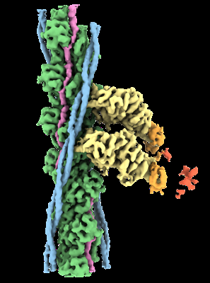 emd_13991.png
 http://ftp.pdbj.org/pub/emdb/structures/EMD-13991


 Z (Sec.)
 Y (Row.)
 X (Col.)


 Sample components
 Processing
 Electron microscopy
 FIELD EMISSION GUN