 Movie
Movie Controller

[English] 日本語
 Yorodumi
- EMDB-13993: In situ structure of myosin neck domain in skeletal sarcomere (ce... -
+ Open data

- Basic information
| Entry | Database: EMDB / ID: EMD-13993 | ||||||||||||||||||
|---|---|---|---|---|---|---|---|---|---|---|---|---|---|---|---|---|---|---|---|
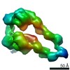
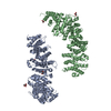
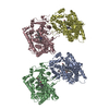
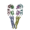
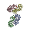
| Title | In situ structure of myosin neck domain in skeletal sarcomere (centered on regulatory light chain) | ||||||||||||||||||
 Map data Map data | |||||||||||||||||||
 Sample Sample |
| ||||||||||||||||||
 Keywords Keywords | Skeletal muscle / Sarcomere / Cross-bridge / MOTOR PROTEIN | ||||||||||||||||||
| Function / homology |  Function and homology information Function and homology informationStriated Muscle Contraction / unconventional myosin complex / contractile muscle fiber / Smooth Muscle Contraction / myosin filament / muscle filament sliding / myosin complex / myosin II complex / structural constituent of muscle / response to muscle activity ...Striated Muscle Contraction / unconventional myosin complex / contractile muscle fiber / Smooth Muscle Contraction / myosin filament / muscle filament sliding / myosin complex / myosin II complex / structural constituent of muscle / response to muscle activity / microfilament motor activity / myofibril / cytoskeletal motor activity / skeletal muscle tissue development / cardiac muscle contraction / muscle contraction / actin filament binding / double-stranded RNA binding / calmodulin binding / calcium ion binding / ATP binding / cytoplasm Similarity search - Function | ||||||||||||||||||
| Biological species |  | ||||||||||||||||||
| Method | subtomogram averaging / cryo EM / Resolution: 9.0 Å | ||||||||||||||||||
 Authors Authors | Wang Z / Grange M | ||||||||||||||||||
| Funding support | European Union, 5 items
| ||||||||||||||||||
 Citation Citation | Journal: Science / Year: 2022 Title: Structures from intact myofibrils reveal mechanism of thin filament regulation through nebulin. Authors: Zhexin Wang / Michael Grange / Sabrina Pospich / Thorsten Wagner / Ay Lin Kho / Mathias Gautel / Stefan Raunser /   Abstract: In skeletal muscle, nebulin stabilizes and regulates the length of thin filaments, but the underlying mechanism remains nebulous. In this work, we used cryo-electron tomography and subtomogram ...In skeletal muscle, nebulin stabilizes and regulates the length of thin filaments, but the underlying mechanism remains nebulous. In this work, we used cryo-electron tomography and subtomogram averaging to reveal structures of native nebulin bound to thin filaments within intact sarcomeres. This in situ reconstruction provided high-resolution details of the interaction between nebulin and actin, demonstrating the stabilizing role of nebulin. Myosin bound to the thin filaments exhibited different conformations of the neck domain, highlighting its inherent structural variability in muscle. Unexpectedly, nebulin did not interact with myosin or tropomyosin, but it did interact with a troponin T linker through two potential binding motifs on nebulin, explaining its regulatory role. Our structures support the role of nebulin as a thin filament "molecular ruler" and provide a molecular basis for studying nemaline myopathies. | ||||||||||||||||||
| History |
|
- Structure visualization
| Movie |
Movie viewer |
|---|---|
| Structure viewer | EM map: SurfViewMolmilJmol/JSmol |
| Supplemental images |

- Downloads & links
-EMDB archive
| Map data | emd_13993.map.gz | 153.8 KB | EMDB map data format | |
|---|---|---|---|---|
| Header (meta data) | emd-13993-v30.xmlemd-13993.xml | 19.8 KB 19.8 KB | Display Display | EMDB header |
| FSC (resolution estimation) | emd_13993_fsc.xml | 3.8 KB | Display | FSC data file |
| Images | 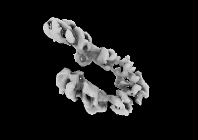 emd_13993.png emd_13993.png | 27.5 KB | ||
| Masks | emd_13993_msk_1.map | 2 MB | Mask map | |
| Filedesc metadata | emd-13993.cif.gz | 6.4 KB | ||
| Others | emd_13993_half_map_1.map.gzemd_13993_half_map_2.map.gz | 1.4 MB 1.4 MB | ||
| Archive directory |  http://ftp.pdbj.org/pub/emdb/structures/EMD-13993ftp://ftp.pdbj.org/pub/emdb/structures/EMD-13993 http://ftp.pdbj.org/pub/emdb/structures/EMD-13993ftp://ftp.pdbj.org/pub/emdb/structures/EMD-13993 | HTTPS FTP |
-Related structure data
| Related structure data |  7qioMC  7qimC  7qinC C: citing same article ( M: atomic model generated by this map |
|---|---|
| Similar structure data |


-Links
| EMDB pages | EMDB (EBI/PDBe) / EMDataResource |
|---|---|
| Related items in Molecule of the Month |


-Map
| File | Download / File: emd_13993.map.gz / Format: CCP4 / Size: 2 MB / Type: IMAGE STORED AS FLOATING POINT NUMBER (4 BYTES) | ||||||||||||||||||||||||||||||||||||||||||||||||||||||||||||
|---|---|---|---|---|---|---|---|---|---|---|---|---|---|---|---|---|---|---|---|---|---|---|---|---|---|---|---|---|---|---|---|---|---|---|---|---|---|---|---|---|---|---|---|---|---|---|---|---|---|---|---|---|---|---|---|---|---|---|---|---|---|
| Projections & slices | Image control
Images are generated by Spider. | ||||||||||||||||||||||||||||||||||||||||||||||||||||||||||||
| Voxel size | X=Y=Z: 3.46 Å | ||||||||||||||||||||||||||||||||||||||||||||||||||||||||||||
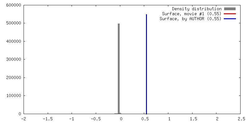
| Density |
| ||||||||||||||||||||||||||||||||||||||||||||||||||||||||||||
| Symmetry | Space group: 1 | ||||||||||||||||||||||||||||||||||||||||||||||||||||||||||||
| Details | EMDB XML:
CCP4 map header:
| ||||||||||||||||||||||||||||||||||||||||||||||||||||||||||||
 Z (Sec.)
 Y (Row.)
 X (Col.)


-Supplemental data
-Mask #1
| File | emd_13993_msk_1.map | ||||||||||||
|---|---|---|---|---|---|---|---|---|---|---|---|---|---|
| Projections & Slices |
| ||||||||||||
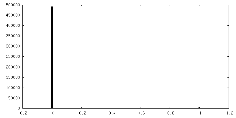
| Density Histograms |


-Half map: #2
| File | emd_13993_half_map_1.map | ||||||||||||
|---|---|---|---|---|---|---|---|---|---|---|---|---|---|
| Projections & Slices |
| ||||||||||||
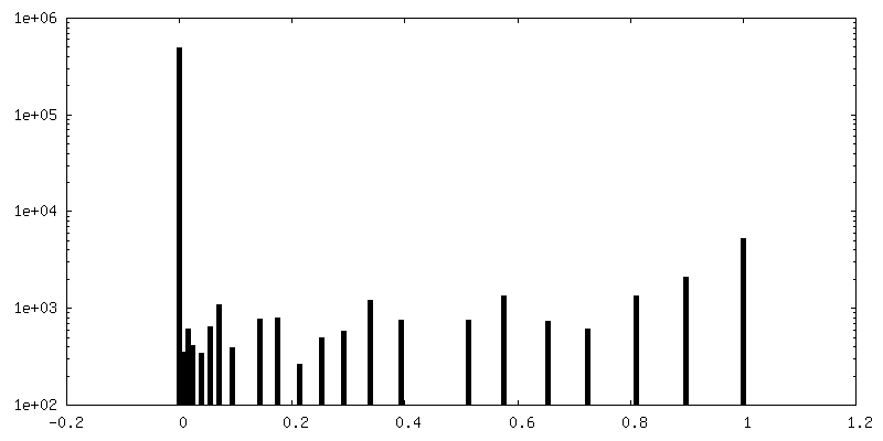
| Density Histograms |


-Half map: #1
| File | emd_13993_half_map_2.map | ||||||||||||
|---|---|---|---|---|---|---|---|---|---|---|---|---|---|
| Projections & Slices |
| ||||||||||||
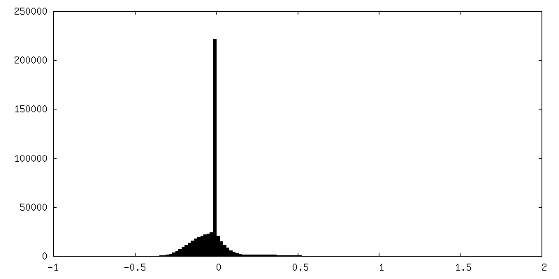
| Density Histograms |


- Sample components
-Entire : Mouse psoas muscle myofibrils
| Entire | Name: Mouse psoas muscle myofibrils |
|---|---|
| Components |
|
-Supramolecule #1: Mouse psoas muscle myofibrils
| Supramolecule | Name: Mouse psoas muscle myofibrils / type: organelle_or_cellular_component / ID: 1 / Parent: 0 / Macromolecule list: all |
|---|---|
| Source (natural) | Organism: |
-Macromolecule #1: Myosin-4
| Macromolecule | Name: Myosin-4 / type: protein_or_peptide / ID: 1 / Number of copies: 2 / Enantiomer: LEVO |
|---|---|
| Source (natural) | Organism: |
| Molecular weight | Theoretical: 96.405609 KDa |
| Sequence | String: MSSDAEMAVF GEAAPYLRKS EKERIEAQNK PFDAKSSVFV VDAKESYVKA TVQSREGGKV TAKTEGGATV TVKDDQVFSM NPPKYDKIE DMAMMTHLHE PAVLYNLKER YAAWMIYTYS GLFCVTVNPY KWLPVYNPEV VAAYRGKKRQ EAPPHIFSIS D NAYQFMLT ...String: MSSDAEMAVF GEAAPYLRKS EKERIEAQNK PFDAKSSVFV VDAKESYVKA TVQSREGGKV TAKTEGGATV TVKDDQVFSM NPPKYDKIE DMAMMTHLHE PAVLYNLKER YAAWMIYTYS GLFCVTVNPY KWLPVYNPEV VAAYRGKKRQ EAPPHIFSIS D NAYQFMLT DRENQSILIT GESGAGKTVN TKRVIQYFAT IAVTGDKKKE EATSGKMQGT LEDQIISANP LLEAFGNAKT VR NDNSSRF GKFIRIHFGA TGKLASADIE TYLLEKSRVT FQLKAERSYH IFYQIMSNKK PELIEMLLIT TNPYDFAYVS QGE ITVPSI DDQEELMATD TAVDILGFSA DEKVAIYKLT GAVMHYGNMK FKQKQREEQA EPDGTEVADK AAYLTSLNSA DLLK ALCYP RVKVGNEYVT KGQTVQQVYN SVGALAKSMY EKMFLWMVTR INQQLDTKQP RQYFIGVLDI AGFEIFDFNT LEQLC INFT NEKLQQFFNH HMFVLEQEEY KKEGIDWEFI DFGMDLAACI ELIEKPMGIF SILEEECMFP KATDTSFKNK LYEQHL GKS NNFQKPKPAK GKAEAHFSLV HYAGTVDYNI IGWLDKNKDP LNETVVGLYQ KSGLKTLAFL FSGGQAAEAE GGGGKKG GK KKGSSFQTVS ALFRENLNKL MTNLKSTHPH FVRCLIPNET KTPGAMEHEL VLHQLRCNGV LEGIRICRKG FPSRILYA D FKQRYKVLNA SAIPEGQFID SKKASEKLLG SIDIDHTQYK FGHTKVFFKA GLLGTLEEMR DEKLAQLITR TQAVCRGYL MRVEFKKMME RRESIFCIQY NVRAFMNVKH WPWMKLYFKI KPLLKSAE UniProtKB: Myosin-4 |
-Macromolecule #2: Myosin light chain 1/3, skeletal muscle isoform
| Macromolecule | Name: Myosin light chain 1/3, skeletal muscle isoform / type: protein_or_peptide / ID: 2 / Number of copies: 2 / Enantiomer: LEVO |
|---|---|
| Source (natural) | Organism: |
| Molecular weight | Theoretical: 20.62049 KDa |
| Sequence | String: MAPKKDVKKP AAAPAPAPAP APAPAKPKEE KIDLSAIKIE FSKEQQEDFK EAFLLFDRTG ECKITLSQVG DVLRALGTNP TNAEVKKVL GNPSNEEMNA KKIEFEQFLP MMQAISNNKD QGGYEDFVEG LRVFDKEGNG TVMGAELRHV LATLGEKMKE E EVEALLAG QEDSNGCINY EAFVKHIMSV UniProtKB: Myosin light chain 1/3, skeletal muscle isoform |
-Macromolecule #3: Myosin regulatory light chain 2, skeletal muscle isoform
| Macromolecule | Name: Myosin regulatory light chain 2, skeletal muscle isoform type: protein_or_peptide / ID: 3 / Number of copies: 2 / Enantiomer: LEVO |
|---|---|
| Source (natural) | Organism: |
| Molecular weight | Theoretical: 18.978445 KDa |
| Sequence | String: MAPKKAKRRA GAEGSSNVFS MFDQTQIQEF KEAFTVIDQN RDGIIDKEDL RDTFAAMGRL NVKNEELDAM MKEASGPINF TVFLTMFGE KLKGADPEDV ITGAFKVLDP EGKGTIKKQF LEELLTTQCD RFSQEEIKNM WAAFPPDVGG NVDYKNICYV I THGDAKDQ E UniProtKB: Myosin regulatory light chain 11 |
-Experimental details
-Structure determination
| Method | cryo EM |
|---|---|
 Processing Processing | subtomogram averaging |
| Aggregation state | tissue |
-Sample preparation
| Buffer | pH: 7 |
|---|---|
| Vitrification | Cryogen name: ETHANE / Instrument: FEI VITROBOT MARK IV |
- Electron microscopy
| Microscope | FEI TITAN KRIOS |
|---|---|
| Image recording | Film or detector model: GATAN K2 SUMMIT (4k x 4k) / Detector mode: COUNTING / Average electron dose: 3.4 e/Å2 |
| Electron beam | Acceleration voltage: 300 kV / Electron source:  FIELD EMISSION GUN FIELD EMISSION GUN |
| Electron optics | C2 aperture diameter: 50.0 µm / Calibrated defocus max: 5.0 µm / Calibrated defocus min: 2.4 µm / Illumination mode: FLOOD BEAM / Imaging mode: BRIGHT FIELD / Cs: 2.7 mm / Nominal defocus max: 5.0 µm / Nominal defocus min: 2.4 µm / Nominal magnification: 81000 |
| Sample stage | Specimen holder model: FEI TITAN KRIOS AUTOGRID HOLDER / Cooling holder cryogen: NITROGEN |
| Experimental equipment |  Model: Titan Krios / Image courtesy: FEI Company |
-Image processing
| Final reconstruction | Applied symmetry - Point group: C1 (asymmetric) / Resolution.type: BY AUTHOR / Resolution: 9.0 Å / Resolution method: FSC 0.143 CUT-OFF / Number subtomograms used: 44001 |
|---|---|
| Extraction | Number tomograms: 48 / Number images used: 183260 |
| Final angle assignment | Type: MAXIMUM LIKELIHOOD |
| FSC plot (resolution estimation) | 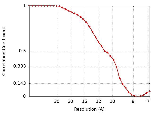 |
-Atomic model buiding 1
| Initial model |
| ||||||||
|---|---|---|---|---|---|---|---|---|---|
| Refinement | Space: REAL / Protocol: RIGID BODY FIT / Overall B value: 300 | ||||||||
| Output model | PDB-7qio: |

