Movie
Movie Controller


+ Open data

- Basic information
| Entry | Database: PDB / ID: 7qin | |||||||||||||||||||||||||||||||||||||||||||||
|---|---|---|---|---|---|---|---|---|---|---|---|---|---|---|---|---|---|---|---|---|---|---|---|---|---|---|---|---|---|---|---|---|---|---|---|---|---|---|---|---|---|---|---|---|---|---|
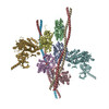
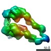
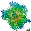
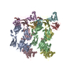
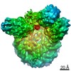
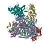
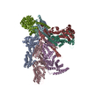
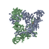
| Title | In situ structure of actomyosin complex in skeletal sarcomere | |||||||||||||||||||||||||||||||||||||||||||||
 Components Components |
| |||||||||||||||||||||||||||||||||||||||||||||
 Keywords Keywords | CONTRACTILE PROTEIN / Skeletal muscle / Sarcomere / Thin filament / Cross-bridge | |||||||||||||||||||||||||||||||||||||||||||||
| Function / homology |  Function and homology information Function and homology informationFormation of the dystrophin-glycoprotein complex (DGC) / Striated Muscle Contraction / Regulation of CDH1 Function / myosin filament / myosin II complex / response to muscle activity / microfilament motor activity / mesenchyme migration / myofibril / striated muscle thin filament ...Formation of the dystrophin-glycoprotein complex (DGC) / Striated Muscle Contraction / Regulation of CDH1 Function / myosin filament / myosin II complex / response to muscle activity / microfilament motor activity / mesenchyme migration / myofibril / striated muscle thin filament / skeletal muscle thin filament assembly / skeletal muscle fiber development / stress fiber / muscle contraction / sarcomere / actin filament / filopodium / Hydrolases; Acting on acid anhydrides; Acting on acid anhydrides to facilitate cellular and subcellular movement / structural constituent of cytoskeleton / actin filament binding / actin cytoskeleton / lamellipodium / double-stranded RNA binding / cell body / calmodulin binding / hydrolase activity / positive regulation of gene expression / protein-containing complex / ATP binding / cytoplasm Similarity search - Function | |||||||||||||||||||||||||||||||||||||||||||||
| Biological species |  | |||||||||||||||||||||||||||||||||||||||||||||
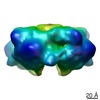
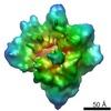
| Method | ELECTRON MICROSCOPY / subtomogram averaging / cryo EM / Resolution: 6.6 Å | |||||||||||||||||||||||||||||||||||||||||||||
 Authors Authors | Wang, Z. / Grange, M. / Pospich, S. / Wagner, T. / Kho, A.L. / Gautel, M. / Raunser, S. | |||||||||||||||||||||||||||||||||||||||||||||
| Funding support | European Union, 5items
| |||||||||||||||||||||||||||||||||||||||||||||
 Citation Citation | Journal: Science / Year: 2022 Title: Structures from intact myofibrils reveal mechanism of thin filament regulation through nebulin. Authors: Zhexin Wang / Michael Grange / Sabrina Pospich / Thorsten Wagner / Ay Lin Kho / Mathias Gautel / Stefan Raunser /   Abstract: In skeletal muscle, nebulin stabilizes and regulates the length of thin filaments, but the underlying mechanism remains nebulous. In this work, we used cryo-electron tomography and subtomogram ...In skeletal muscle, nebulin stabilizes and regulates the length of thin filaments, but the underlying mechanism remains nebulous. In this work, we used cryo-electron tomography and subtomogram averaging to reveal structures of native nebulin bound to thin filaments within intact sarcomeres. This in situ reconstruction provided high-resolution details of the interaction between nebulin and actin, demonstrating the stabilizing role of nebulin. Myosin bound to the thin filaments exhibited different conformations of the neck domain, highlighting its inherent structural variability in muscle. Unexpectedly, nebulin did not interact with myosin or tropomyosin, but it did interact with a troponin T linker through two potential binding motifs on nebulin, explaining its regulatory role. Our structures support the role of nebulin as a thin filament "molecular ruler" and provide a molecular basis for studying nemaline myopathies. | |||||||||||||||||||||||||||||||||||||||||||||
| History |
|
- Structure visualization
| Movie |
Movie viewer |
|---|---|
| Structure viewer | Molecule: MolmilJmol/JSmol |
- Downloads & links
-Download
| PDBx/mmCIF format | 7qin.cif.gz | 642.9 KB | Display | PDBx/mmCIF format |
|---|---|---|---|---|
| PDB format | pdb7qin.ent.gz | 533.1 KB | Display | PDB format |
| PDBx/mmJSON format | 7qin.json.gz | Tree view | PDBx/mmJSON format | |
| Others |  Other downloads Other downloads |
-Validation report
| Arichive directory | https://data.pdbj.org/pub/pdb/validation_reports/qi/7qinftp://data.pdbj.org/pub/pdb/validation_reports/qi/7qin | HTTPS FTP |
|---|
-Related structure data
| Related structure data |  13991MC  7qimC  7qioC C: citing same article ( M: map data used to model this data |
|---|---|
| Similar structure data |


-Links
 PDBj
PDBj


- Assembly
| Deposited unit | 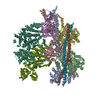
|
|---|---|
| 1 |
|
-Components
-Protein , 4 types, 9 molecules ABCDEFGLM
| #1: Protein | Mass: 41387.227 Da / Num. of mol.: 5 / Source method: isolated from a natural source / Source: (natural) #2: Protein | | Mass: 9188.235 Da / Num. of mol.: 1 / Source method: isolated from a natural source Details: Model with conserved tyrosines of three nebulin simple repeats Source: (natural) #3: Protein | | Mass: 6131.495 Da / Num. of mol.: 1 / Source method: isolated from a natural source Details: Model with conserved tyrosines of two nebulin simple repeats Source: (natural) #7: Protein | Mass: 96405.609 Da / Num. of mol.: 2 / Source method: isolated from a natural source / Source: (natural) |
|---|
-Tropomyosin, alpha-1 ... , 3 types, 4 molecules HIJK
| #4: Protein | Mass: 9975.288 Da / Num. of mol.: 1 / Source method: isolated from a natural source / Source: (natural) |
|---|---|
| #5: Protein | Mass: 10060.393 Da / Num. of mol.: 1 / Source method: isolated from a natural source / Source: (natural) |
| #6: Protein | Mass: 7507.245 Da / Num. of mol.: 2 / Source method: isolated from a natural source / Source: (natural) |
-Non-polymers , 2 types, 10 molecules 

| #8: Chemical | ChemComp-ADP /  Mass: 427.201 Da / Num. of mol.: 5 / Source method: obtained synthetically / Formula: C10H15N5O10P2 / Comment: ADP, energy-carrying molecule*YM Mass: 427.201 Da / Num. of mol.: 5 / Source method: obtained synthetically / Formula: C10H15N5O10P2 / Comment: ADP, energy-carrying molecule*YM#9: Chemical | ChemComp-MG /  Mass: 24.305 Da / Num. of mol.: 5 / Source method: obtained synthetically / Formula: Mg Mass: 24.305 Da / Num. of mol.: 5 / Source method: obtained synthetically / Formula: Mg |
|---|
-Details
| Has ligand of interest | N |
|---|---|
| Has protein modification | Y |
-Experimental details
-Experiment
| Experiment | Method: ELECTRON MICROSCOPY |
|---|---|
| EM experiment | Aggregation state: TISSUE / 3D reconstruction method: subtomogram averaging |
- Sample preparation
| Component | Name: Mouse psoas muscle myofibrils / Type: ORGANELLE OR CELLULAR COMPONENT / Entity ID: #1-#7 / Source: NATURAL |
|---|---|
| Source (natural) | Organism: |
| Buffer solution | pH: 7 |
| Specimen | Embedding applied: NO / Shadowing applied: NO / Staining applied: NO / Vitrification applied: YES |
| Vitrification | Instrument: FEI VITROBOT MARK IV / Cryogen name: ETHANE |
- Electron microscopy imaging
| Experimental equipment |  Model: Titan Krios / Image courtesy: FEI Company |
|---|---|
| Microscopy | Model: FEI TITAN KRIOS |
| Electron gun | Electron source:  FIELD EMISSION GUN / Accelerating voltage: 300 kV / Illumination mode: FLOOD BEAM FIELD EMISSION GUN / Accelerating voltage: 300 kV / Illumination mode: FLOOD BEAM |
| Electron lens | Mode: BRIGHT FIELD / Nominal magnification: 81000 X / Calibrated defocus min: 2400 nm / Calibrated defocus max: 5000 nm / Cs: 2.7 mm / C2 aperture diameter: 50 µm |
| Specimen holder | Cryogen: NITROGEN / Specimen holder model: FEI TITAN KRIOS AUTOGRID HOLDER |
| Image recording | Electron dose: 3.4 e/Å2 / Detector mode: COUNTING / Film or detector model: GATAN K2 SUMMIT (4k x 4k) |
- Processing
| EM software |
| |||||||||||||||
|---|---|---|---|---|---|---|---|---|---|---|---|---|---|---|---|---|
| CTF correction | Type: PHASE FLIPPING AND AMPLITUDE CORRECTION | |||||||||||||||
| Symmetry | Point symmetry: C1 (asymmetric) | |||||||||||||||
| 3D reconstruction | Resolution: 6.6 Å / Resolution method: FSC 0.143 CUT-OFF / Num. of particles: 104302 / Symmetry type: POINT | |||||||||||||||
| EM volume selection | Num. of tomograms: 48 / Num. of volumes extracted: 183260 | |||||||||||||||
| Atomic model building | B value: 75 / Space: REAL Details: Mg was not refined but copied from the initial model | |||||||||||||||
| Atomic model building | 3D fitting-ID: 1 / Pdb chain-ID: A / Source name: PDB / Type: experimental model
|