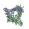
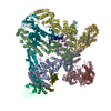
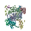
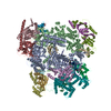
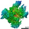
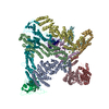
Entry Database : PDB / ID : 5cslTitle Crystal structure of the 500 kD yeast acetyl-CoA carboxylase holoenzyme dimer Acetyl-CoA carboxylase Keywords / Function / homology Function Domain/homology Component
/ / / / / / / / / / / / / / / / / / / / / / / / / / / / / / / / / / / / / / / / / / / / / / / / / / / / / / / / / / / / / / / / / / / / / / / / / / / / / / / / / / Biological species Saccharomyces cerevisiae (brewer's yeast)Method / / / / Resolution : 3.2 Å Authors Wei, J. / Tong, L. Funding support Organization Grant number Country National Institutes of Health/Office of the Director OD012018 National Institutes of Health/National Institute of Diabetes and Digestive and Kidney Disease (NIH/NIDDK) DK067238
Journal : Nature / Year : 2015Title : Crystal structure of the 500-kDa yeast acetyl-CoA carboxylase holoenzyme dimer.Authors : Wei, J. / Tong, L. History Deposition Jul 23, 2015 Deposition site / Processing site Revision 1.0 Oct 28, 2015 Provider / Type Revision 1.1 Nov 11, 2015 Group Revision 1.2 Sep 20, 2017 Group / Derived calculations / Category / pdbx_struct_oper_listItem / _pdbx_struct_oper_list.symmetry_operationRevision 1.3 Mar 23, 2022 Group / Database references / Category / pdbx_audit_supportItem _database_2.pdbx_DOI / _database_2.pdbx_database_accession ... _database_2.pdbx_DOI / _database_2.pdbx_database_accession / _pdbx_audit_support.funding_organization / _pdbx_audit_support.grant_number Revision 1.4 Mar 4, 2026 Group / Refinement description / Structure summaryCategory chem_comp_atom / chem_comp_bond ... chem_comp_atom / chem_comp_bond / pdbx_entry_details / pdbx_initial_refinement_model
Show all Show less
 Movie
Movie Controller

 Yorodumi
 Open data
 Basic information
 Components
 Keywords
 Function and homology information
 X-RAY DIFFRACTION /
  Authors
 United States, 2items
  Citation
 Structure visualization
 Downloads & links
 Other downloads


 PDBj
PDBj


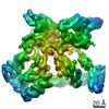
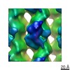
 Assembly
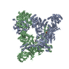


 Mass: 228.311 Da / Num. of mol.: 2 / Source method: obtained synthetically / Formula: C10H16N2O2S
 Mass: 767.534 Da / Num. of mol.: 2 / Source method: obtained synthetically / Formula: C21H36N7O16P3S
 Sample preparation
 Processing