 Movie
Movie Controller

[English] 日本語
 Yorodumi
- PDB-7pog: High-resolution structure of native toxin A from Clostridioides d... -
+ Open data

- Basic information
| Entry | Database: PDB / ID: 7pog | ||||||
|---|---|---|---|---|---|---|---|
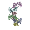
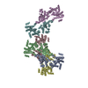
| Title | High-resolution structure of native toxin A from Clostridioides difficile | ||||||
 Components Components | Toxin A | ||||||
 Keywords Keywords | TOXIN / glucosyltransferase | ||||||
| Function / homology |  Function and homology information Function and homology informationTransferases; Glycosyltransferases; Hexosyltransferases / host cell cytosol / glycosyltransferase activity / cysteine-type peptidase activity / host cell endosome membrane / toxin activity / lipid binding / host cell plasma membrane / proteolysis / extracellular region / metal ion binding Similarity search - Function | ||||||
| Biological species |  Clostridioides difficile (bacteria) Clostridioides difficile (bacteria) | ||||||
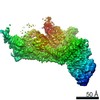
| Method | ELECTRON MICROSCOPY / single particle reconstruction / cryo EM / Resolution: 2.83 Å | ||||||
 Authors Authors | Boesen, T. / Joergensen, R. / Aminzadeh, A. / Engelbrecht Larsen, C. | ||||||
| Funding support |  Denmark, 1items Denmark, 1items
| ||||||
 Citation Citation | Journal: EMBO Rep / Year: 2022 Title: High-resolution structure of native toxin A from Clostridioides difficile. Authors: Aria Aminzadeh / Christian Engelbrecht Larsen / Thomas Boesen / René Jørgensen / Abstract: Clostridioides difficile infections have emerged as the leading cause of healthcare-associated infectious diarrhea. Disease symptoms are mainly caused by the virulence factors, TcdA and TcdB, which ...Clostridioides difficile infections have emerged as the leading cause of healthcare-associated infectious diarrhea. Disease symptoms are mainly caused by the virulence factors, TcdA and TcdB, which are large homologous multidomain proteins. Here, we report a 2.8 Å resolution cryo-EM structure of native TcdA, unveiling its conformation at neutral pH. The structure uncovers the dynamic movement of the CROPs domain which is induced in response to environmental acidification. Furthermore, the structure reveals detailed information about the interaction area between the CROPs domain and the tip of the delivery and receptor-binding domain, which likely serves to shield the C-terminal part of the hydrophobic pore-forming region from solvent exposure. Similarly, extensive interactions between the globular subdomain and the N-terminal part of the pore-forming region suggest that the globular subdomain shields the upper part of the pore-forming region from exposure to the surrounding solvent. Hence, the TcdA structure provides insights into the mechanism of preventing premature unfolding of the pore-forming region at neutral pH, as well as the pH-induced inter-domain dynamics. | ||||||
| History |
|
- Structure visualization
| Movie |
Movie viewer |
|---|---|
| Structure viewer | Molecule: MolmilJmol/JSmol |
- Downloads & links
-Download
| PDBx/mmCIF format | 7pog.cif.gz | 433.6 KB | Display | PDBx/mmCIF format |
|---|---|---|---|---|
| PDB format | pdb7pog.ent.gz | 346.1 KB | Display | PDB format |
| PDBx/mmJSON format | 7pog.json.gz | Tree view | PDBx/mmJSON format | |
| Others |  Other downloads Other downloads |
-Validation report
| Arichive directory | https://data.pdbj.org/pub/pdb/validation_reports/po/7pogftp://data.pdbj.org/pub/pdb/validation_reports/po/7pog | HTTPS FTP |
|---|
-Related structure data
| Related structure data |  13574MC M: map data used to model this data C: citing same article ( |
|---|---|
| Similar structure data |


-Links
 PDBj
PDBj


- Assembly
| Deposited unit | 
|
|---|---|
| 1 |
|
-Components
| #1: Protein | Mass: 308613.188 Da / Num. of mol.: 1 / Source method: isolated from a natural source / Source: (natural) Clostridioides difficile (bacteria) / References: UniProt: M4NKU0 |
|---|---|
| #2: Chemical | ChemComp-ZN /   Mass: 65.409 Da / Num. of mol.: 1 / Source method: obtained synthetically / Formula: Zn / Feature type: SUBJECT OF INVESTIGATION Mass: 65.409 Da / Num. of mol.: 1 / Source method: obtained synthetically / Formula: Zn / Feature type: SUBJECT OF INVESTIGATION |
| Has ligand of interest | Y |
-Experimental details
-Experiment
| Experiment | Method: ELECTRON MICROSCOPY |
|---|---|
| EM experiment | Aggregation state: PARTICLE / 3D reconstruction method: single particle reconstruction |
- Sample preparation
| Component | Name: Clostridioides difficile Toxin A / Type: ORGANELLE OR CELLULAR COMPONENT / Entity ID: #1 / Source: NATURAL |
|---|---|
| Source (natural) | Organism: Clostridioides difficile (bacteria) |
| Buffer solution | pH: 7.5 |
| Specimen | Embedding applied: NO / Shadowing applied: NO / Staining applied: NO / Vitrification applied: YES |
| Vitrification | Cryogen name: ETHANE |
- Electron microscopy imaging
| Experimental equipment |  Model: Titan Krios / Image courtesy: FEI Company |
|---|---|
| Microscopy | Model: FEI TITAN KRIOS |
| Electron gun | Electron source:  FIELD EMISSION GUN / Accelerating voltage: 300 kV / Illumination mode: FLOOD BEAM FIELD EMISSION GUN / Accelerating voltage: 300 kV / Illumination mode: FLOOD BEAM |
| Electron lens | Mode: BRIGHT FIELD |
| Image recording | Electron dose: 60 e/Å2 / Film or detector model: GATAN K3 BIOQUANTUM (6k x 4k) |
- Processing
| CTF correction | Type: NONE |
|---|---|
| 3D reconstruction | Resolution: 2.83 Å / Resolution method: FSC 0.143 CUT-OFF / Num. of particles: 900000 / Symmetry type: POINT |
| Atomic model building | Protocol: AB INITIO MODEL |
