[English] 日本語
 Yorodumi
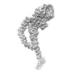
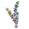
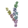
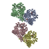
- EMDB-13574: High-resolution structure of native toxin A from Clostridioides d... -
+ Open data
- Basic information
| Entry | Database: EMDB / ID: EMD-13574 | |||||||||
|---|---|---|---|---|---|---|---|---|---|---|
| Title | High-resolution structure of native toxin A from Clostridioides difficile | |||||||||
 Map data Map data | ||||||||||
 Sample Sample |
| |||||||||
 Keywords Keywords | glucosyltransferase / TOXIN | |||||||||
| Function / homology |  Function and homology information Function and homology informationTransferases; Glycosyltransferases; Hexosyltransferases / host cell cytosol / glycosyltransferase activity / cysteine-type peptidase activity / host cell endosome membrane / toxin activity / lipid binding / host cell plasma membrane / proteolysis / extracellular region ...Transferases; Glycosyltransferases; Hexosyltransferases / host cell cytosol / glycosyltransferase activity / cysteine-type peptidase activity / host cell endosome membrane / toxin activity / lipid binding / host cell plasma membrane / proteolysis / extracellular region / metal ion binding / membrane Similarity search - Function | |||||||||
| Biological species |  Clostridioides difficile (bacteria) Clostridioides difficile (bacteria) | |||||||||
| Method | single particle reconstruction / cryo EM / Resolution: 2.83 Å | |||||||||
 Authors Authors | Boesen T / Joergensen R | |||||||||
| Funding support |  Denmark, 1 items Denmark, 1 items
| |||||||||
 Citation Citation |  Journal: EMBO Rep / Year: 2022 Journal: EMBO Rep / Year: 2022Title: High-resolution structure of native toxin A from Clostridioides difficile. Authors: Aria Aminzadeh / Christian Engelbrecht Larsen / Thomas Boesen / René Jørgensen /  Abstract: Clostridioides difficile infections have emerged as the leading cause of healthcare-associated infectious diarrhea. Disease symptoms are mainly caused by the virulence factors, TcdA and TcdB, which ...Clostridioides difficile infections have emerged as the leading cause of healthcare-associated infectious diarrhea. Disease symptoms are mainly caused by the virulence factors, TcdA and TcdB, which are large homologous multidomain proteins. Here, we report a 2.8 Å resolution cryo-EM structure of native TcdA, unveiling its conformation at neutral pH. The structure uncovers the dynamic movement of the CROPs domain which is induced in response to environmental acidification. Furthermore, the structure reveals detailed information about the interaction area between the CROPs domain and the tip of the delivery and receptor-binding domain, which likely serves to shield the C-terminal part of the hydrophobic pore-forming region from solvent exposure. Similarly, extensive interactions between the globular subdomain and the N-terminal part of the pore-forming region suggest that the globular subdomain shields the upper part of the pore-forming region from exposure to the surrounding solvent. Hence, the TcdA structure provides insights into the mechanism of preventing premature unfolding of the pore-forming region at neutral pH, as well as the pH-induced inter-domain dynamics. | |||||||||
| History |
|
- Structure visualization
| Movie |
 Movie viewer Movie viewer |
|---|---|
| Structure viewer | EM map:  SurfView SurfView Molmil Molmil Jmol/JSmol Jmol/JSmol |
| Supplemental images |
- Downloads & links
-EMDB archive
| Map data |  emd_13574.map.gz emd_13574.map.gz | 534.7 MB |  EMDB map data format EMDB map data format | |
|---|---|---|---|---|
| Header (meta data) |  emd-13574-v30.xml emd-13574-v30.xml emd-13574.xml emd-13574.xml | 11.4 KB 11.4 KB | Display Display |  EMDB header EMDB header |
| Images |  emd_13574.png emd_13574.png | 69.1 KB | ||
| Filedesc metadata |  emd-13574.cif.gz emd-13574.cif.gz | 6 KB | ||
| Archive directory |  http://ftp.pdbj.org/pub/emdb/structures/EMD-13574 http://ftp.pdbj.org/pub/emdb/structures/EMD-13574 ftp://ftp.pdbj.org/pub/emdb/structures/EMD-13574 ftp://ftp.pdbj.org/pub/emdb/structures/EMD-13574 | HTTPS FTP |
-Validation report
| Summary document |  emd_13574_validation.pdf.gz emd_13574_validation.pdf.gz | 481.7 KB | Display |  EMDB validaton report EMDB validaton report |
|---|---|---|---|---|
| Full document |  emd_13574_full_validation.pdf.gz emd_13574_full_validation.pdf.gz | 481.3 KB | Display | |
| Data in XML |  emd_13574_validation.xml.gz emd_13574_validation.xml.gz | 9 KB | Display | |
| Data in CIF |  emd_13574_validation.cif.gz emd_13574_validation.cif.gz | 10.4 KB | Display | |
| Arichive directory |  https://ftp.pdbj.org/pub/emdb/validation_reports/EMD-13574 https://ftp.pdbj.org/pub/emdb/validation_reports/EMD-13574 ftp://ftp.pdbj.org/pub/emdb/validation_reports/EMD-13574 ftp://ftp.pdbj.org/pub/emdb/validation_reports/EMD-13574 | HTTPS FTP |
-Related structure data
| Related structure data |  7pogMC M: atomic model generated by this map C: citing same article ( |
|---|---|
| Similar structure data |
- Links
Links
| EMDB pages |  EMDB (EBI/PDBe) / EMDB (EBI/PDBe) /  EMDataResource EMDataResource |
|---|---|
| Related items in Molecule of the Month |
- Map
Map
| File |  Download / File: emd_13574.map.gz / Format: CCP4 / Size: 1.1 GB / Type: IMAGE STORED AS FLOATING POINT NUMBER (4 BYTES) Download / File: emd_13574.map.gz / Format: CCP4 / Size: 1.1 GB / Type: IMAGE STORED AS FLOATING POINT NUMBER (4 BYTES) | ||||||||||||||||||||||||||||||||||||||||||||||||||||||||||||
|---|---|---|---|---|---|---|---|---|---|---|---|---|---|---|---|---|---|---|---|---|---|---|---|---|---|---|---|---|---|---|---|---|---|---|---|---|---|---|---|---|---|---|---|---|---|---|---|---|---|---|---|---|---|---|---|---|---|---|---|---|---|
| Projections & slices | Image control
Images are generated by Spider. | ||||||||||||||||||||||||||||||||||||||||||||||||||||||||||||
| Voxel size | X=Y=Z: 0.64 Å | ||||||||||||||||||||||||||||||||||||||||||||||||||||||||||||
| Density |
| ||||||||||||||||||||||||||||||||||||||||||||||||||||||||||||
| Symmetry | Space group: 1 | ||||||||||||||||||||||||||||||||||||||||||||||||||||||||||||
| Details | EMDB XML:
CCP4 map header:
| ||||||||||||||||||||||||||||||||||||||||||||||||||||||||||||
-Supplemental data
- Sample components
-Entire : Clostridioides difficile Toxin A
| Entire | Name: Clostridioides difficile Toxin A |
|---|---|
| Components |
|
-Supramolecule #1: Clostridioides difficile Toxin A
| Supramolecule | Name: Clostridioides difficile Toxin A / type: organelle_or_cellular_component / ID: 1 / Parent: 0 / Macromolecule list: #1 |
|---|---|
| Source (natural) | Organism:  Clostridioides difficile (bacteria) Clostridioides difficile (bacteria) |
-Macromolecule #1: Toxin A
| Macromolecule | Name: Toxin A / type: protein_or_peptide / ID: 1 / Number of copies: 1 / Enantiomer: LEVO |
|---|---|
| Source (natural) | Organism:  Clostridioides difficile (bacteria) Clostridioides difficile (bacteria) |
| Molecular weight | Theoretical: 308.613188 KDa |
| Sequence | String: MSLISKEELI KLAYSIRPRE NEYKTILTNL DEYNKLTTNN NENKYLQLKK LNESIDVFMN KYKNSSRNRA LSNLKKDILK EVILIKNSN TSPVEKNLHF VWIGGEVSDI ALEYIKQWAD INAEYNIKLW YDSEAFLVNT LKKAIVESST TEALQLLEEE I QNPQFDNM ...String: MSLISKEELI KLAYSIRPRE NEYKTILTNL DEYNKLTTNN NENKYLQLKK LNESIDVFMN KYKNSSRNRA LSNLKKDILK EVILIKNSN TSPVEKNLHF VWIGGEVSDI ALEYIKQWAD INAEYNIKLW YDSEAFLVNT LKKAIVESST TEALQLLEEE I QNPQFDNM KFYKKRMEFI YDRQKRFINY YKSQINKPTV PTIDDIIKSH LVSEYNRDET LLESYRTNSL RKINSNHGID IR ANSLFTE QELLNIYSQE LLNRGNLAAA SDIVRLLALK NFGGVYLDVD MLPGIHSDLF KTIPRPSSIG LDRWEMIKLE AIM KYKKYI NNYTSENFDK LDQQLKDNFK LIIESKSEKS EIFSKLENLN VSDLEIKIAF ALGSVINQAL ISKQGSYLTN LVIE QVKNR YQFLNQHLNP AIESDNNFTD TTKIFHDSLF NSATAENSMF LTKIAPYLQV GFMPEARSTI SLSGPGAYAS AYYDF INLQ ENTIEKTLKA SDLIEFKFPE NNLSQLTEQE INSLWSFDQA SAKYQFEKYV RDYTGGSLSE DNGVDFNKNT ALDKNY LLN NKIPSNNVEE AGSKNYVHYI IQLQGDDISY EATCNLFSKN PKNSIIIQRN MNESAKSYFL SDDGESILEL NKYRIPE RL KNKEKVKVTF IGHGKDEFNT SEFARLSVDS LSNEISSFLD TIKLDISPKN VEVNLLGCNM FSYDFNVEET YPGKLLLS I MDKITSTLPD VNKDSITIGA NQYEVRINSE GRKELLAHSG KWINKEEAIM SDLSSKEYIF FDSIDNKLKA KSKNIPGLA SISEDIKTLL LDASVSPDTK FILNNLKLNI ESSIGDYIYY EKLEPVKNII HNSIDDLIDE FNLLENVSDE LYELKKLNNL DEKYLISFE DISKNNSTYS VRFINKSNGE SVYVETEKEI FSKYSEHITK EISTIKNSII TDVNGNLLDN IQLDHTSQVN T LNAAFFIQ SLIDYSSNKD VLNDLSTSVK VQLYAQLFST GLNTIYDSIQ LVNLISNAVN DTINVLPTIT EGIPIVSTIL DG INLGAAI KELLDEHDPL LKKELEAKVG VLAINMSLSI AATVASIVGI GAEVTIFLLP IAGISAGIPS LVNNELILHD KAT SVVNYF NHLSESKEYG PLKTEDDKIL VPIDDLVISE IDFNNNSIKL GTCNILAMEG GSGHTVTGNI DHFFSSPYIS SHIP SLSVY SAIGIKTENL DFSKKIMMLP NAPSRVFWWE TGAVPGLRSL ENNGTKLLDS IRDLYPGKFY WRFYAFFDYA ITTLK PVYE DTNTKIKLDK DTRNFIMPTI TTDEIRNKLS YSFDGAGGTY SLLLSSYPIS MNINLSKDDL WIFNIDNEVR EISIEN GTI KKGNLIEDVL SKIDINKNKL IIGNQTIDFS GDIDNKDRYI FLTCELDDKI SLIIEINLVA KSYSLLLSGD KNYLISN LS NTIEKINTLG LDSKNIAYNY TDESNNKYFG AISKTSQKSI IHYKKDSKNI LEFYNGSTLE FNSKDFIAED INVFMKDD I NTITGKYYVD NNTDKSIDFS ISLVSKNQVK VNGLYLNESV YSSYLDFVKN SDGHHNTSNF MNLFLNNISF WKLFGFENI NFVIDKYFTL VGKTNLGYVE FICDNNKNID IYFGEWKTSS SKSTIFSGNG RNVVVEPIYN PDTGEDISTS LDFSYEPLYG IDRYINKVL IAPDLYTSLI NINTNYYSNE YYPEIIVLNP NTFHKKVNIN LDSSSFEYKW STEGSDFILV RYLEESNKKI L QKIRIKGI LSNTQSFNKM SIDFKDIKKL SLGYIMSNFK SFNSENELDR DHLGFKIIDN KTYYYDEDSK LVKGLININN SL FYFDPIE SNLVTGWQTI NGKKYYFDIN TGAASTSYKI INGKHFYFNN NGVMQLGVFK GPDGFEYFAP ANTQNNNIEG QAI VYQSKF LTLNGKKYYF DNDSKAVTGW RIINNEKYYF NPNNAIAAVG LQVIDNNKYY FNPDTAIISK GWQTVNGSRY YFDT DTAIA FNGYKTIDGK HFYFDSDCVV KIGVFSGSNG FEYFAPANTY NNNIEGQAIV YQSKFLTLNG KKYYFDNNSK AVTGW QTID SKKYYFNTNT AEAATGWQTI DGKKYYFNTN TAEAATGWQT IDGKKYYFNT NTSIASTGYT IINGKYFYFN TDGIMQ IGV FKVPNGFEYF APANTHNNNI EGQAILYQNK FLTLNGKKYY FGSDSKAITG WQTIDGKKYY FNPNNAIAAT HLCTINN DK YYFSYDGILQ NGYITIERNN FYFDANNESK MVTGVFKGPN GFEYFAPANT HNNNIEGQAI VYQNKFLTLN GKKYYFDN D SKAVTGWQTI DSKKYYFNLN TAVAVTGWQT IDGEKYYFNL NTAEAATGWQ TIDGKRYYFN TNTYIASTGY TIINGKHFY FNTDGIMQIG VFKGPDGFEY FAPANTHNNN IEGQAILYQN KFLTLNGKKY YFGSDSKAVT GLRTIDGKKY YFNTNTAVAV TGWQTINGK KYYFNTNTYI ASTGYTIISG KHFYFNTDGI MQIGVFKGPD GFEYFAPANT DANNIEGQAI RYQNRFLYLH D NIYYFGND SKAATGWATI DGNRYYFEPN TAMGANGYKT IDNKNFYFRN GLPQIGVFKG PNGFEYFAPA NTDANNIDGQ AI RYQNRFL HLLGKIYYFG NNSKAVTGWQ TINSKVYYFM PDTAMAAAGG LFEIDGVIYF FGVDGVKAPG IYG UniProtKB: Toxin A |
-Macromolecule #2: ZINC ION
| Macromolecule | Name: ZINC ION / type: ligand / ID: 2 / Number of copies: 1 / Formula: ZN |
|---|---|
| Molecular weight | Theoretical: 65.409 Da |
-Experimental details
-Structure determination
| Method | cryo EM |
|---|---|
 Processing Processing | single particle reconstruction |
| Aggregation state | particle |
- Sample preparation
| Buffer | pH: 7.5 |
|---|---|
| Vitrification | Cryogen name: ETHANE |
- Electron microscopy
| Microscope | FEI TITAN KRIOS |
|---|---|
| Image recording | Film or detector model: GATAN K3 BIOQUANTUM (6k x 4k) / Average electron dose: 60.0 e/Å2 |
| Electron beam | Acceleration voltage: 300 kV / Electron source:  FIELD EMISSION GUN FIELD EMISSION GUN |
| Electron optics | Illumination mode: FLOOD BEAM / Imaging mode: BRIGHT FIELD |
| Experimental equipment |  Model: Titan Krios / Image courtesy: FEI Company |
- Image processing
| Startup model | Type of model: NONE |
|---|---|
| Final reconstruction | Resolution.type: BY AUTHOR / Resolution: 2.83 Å / Resolution method: FSC 0.143 CUT-OFF / Number images used: 900000 |
| Initial angle assignment | Type: MAXIMUM LIKELIHOOD |
| Final angle assignment | Type: MAXIMUM LIKELIHOOD |
-Atomic model buiding 1
| Refinement | Protocol: AB INITIO MODEL |
|---|---|
| Output model |  PDB-7pog: |
 Movie
Movie Controller


 Z (Sec.)
 Y (Row.)
 X (Col.)


