 Movie
Movie Controller

[English] 日本語
 Yorodumi
- PDB-7p34: Cryo-EM structure of the proton-dependent antibacterial peptide t... -
+ Open data

- Basic information
| Entry | Database: PDB / ID: 7p34 | ||||||||||||||||||
|---|---|---|---|---|---|---|---|---|---|---|---|---|---|---|---|---|---|---|---|
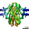
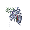
| Title | Cryo-EM structure of the proton-dependent antibacterial peptide transporter SbmA-FabS11-1 in nanodiscs | ||||||||||||||||||
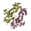
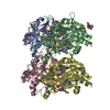
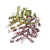
 Components Components | Peptide antibiotic transporter SbmA | ||||||||||||||||||
 Keywords Keywords | TRANSPORT PROTEIN / SLiPT / proton transport / peptide transport / antibiotics | ||||||||||||||||||
| Function / homology |  Function and homology information Function and homology informationsecondary active transmembrane transporter activity / microcin transmembrane transporter activity / microcin B17 transport / : / microcin transport / toxin transmembrane transporter activity / plasma membrane => GO:0005886 / peptide transmembrane transporter activity / peptide transport / ATPase-coupled transmembrane transporter activity ...secondary active transmembrane transporter activity / microcin transmembrane transporter activity / microcin B17 transport / : / microcin transport / toxin transmembrane transporter activity / plasma membrane => GO:0005886 / peptide transmembrane transporter activity / peptide transport / ATPase-coupled transmembrane transporter activity / protein transport / response to antibiotic / ATP hydrolysis activity / ATP binding / plasma membrane Similarity search - Function | ||||||||||||||||||
| Biological species |  | ||||||||||||||||||
| Method | ELECTRON MICROSCOPY / single particle reconstruction / cryo EM / Resolution: 3.59 Å | ||||||||||||||||||
 Authors Authors | Ghilarov, D. / Beis, K. | ||||||||||||||||||
| Funding support |  United Kingdom, United Kingdom,  Japan, Japan,  Poland, 5items Poland, 5items
| ||||||||||||||||||
 Citation Citation | Journal: Sci Adv / Year: 2021 Title: Molecular mechanism of SbmA, a promiscuous transporter exploited by antimicrobial peptides. Authors: Dmitry Ghilarov / Satomi Inaba-Inoue / Piotr Stepien / Feng Qu / Elizabeth Michalczyk / Zuzanna Pakosz / Norimichi Nomura / Satoshi Ogasawara / Graham Charles Walker / Sylvie Rebuffat / So ...Authors: Dmitry Ghilarov / Satomi Inaba-Inoue / Piotr Stepien / Feng Qu / Elizabeth Michalczyk / Zuzanna Pakosz / Norimichi Nomura / Satoshi Ogasawara / Graham Charles Walker / Sylvie Rebuffat / So Iwata / Jonathan Gardiner Heddle / Konstantinos Beis /   Abstract: Antibiotic metabolites and antimicrobial peptides mediate competition between bacterial species. Many of them hijack inner and outer membrane proteins to enter cells. Sensitivity of enteric bacteria ...Antibiotic metabolites and antimicrobial peptides mediate competition between bacterial species. Many of them hijack inner and outer membrane proteins to enter cells. Sensitivity of enteric bacteria to multiple peptide antibiotics is controlled by the single inner membrane protein SbmA. To establish the molecular mechanism of peptide transport by SbmA and related BacA, we determined their cryo–electron microscopy structures at 3.2 and 6 Å local resolution, respectively. The structures show a previously unknown fold, defining a new class of secondary transporters named SbmA-like peptide transporters. The core domain includes conserved glutamates, which provide a pathway for proton translocation, powering transport. The structures show an outward-open conformation with a large cavity that can accommodate diverse substrates. We propose a molecular mechanism for antibacterial peptide uptake paving the way for creation of narrow-targeted therapeutics. | ||||||||||||||||||
| History |
|
- Structure visualization
| Movie |
Movie viewer |
|---|---|
| Structure viewer | Molecule: MolmilJmol/JSmol |
- Downloads & links
-Download
| PDBx/mmCIF format | 7p34.cif.gz | 149.3 KB | Display | PDBx/mmCIF format |
|---|---|---|---|---|
| PDB format | pdb7p34.ent.gz | 118.1 KB | Display | PDB format |
| PDBx/mmJSON format | 7p34.json.gz | Tree view | PDBx/mmJSON format | |
| Others |  Other downloads Other downloads |
-Validation report
| Summary document | 7p34_validation.pdf.gz | 747.8 KB | Display | wwPDB validaton report |
|---|---|---|---|---|
| Full document | 7p34_full_validation.pdf.gz | 753.9 KB | Display | |
| Data in XML | 7p34_validation.xml.gz | 37.2 KB | Display | |
| Data in CIF | 7p34_validation.cif.gz | 50.8 KB | Display | |
| Arichive directory | https://data.pdbj.org/pub/pdb/validation_reports/p3/7p34ftp://data.pdbj.org/pub/pdb/validation_reports/p3/7p34 | HTTPS FTP |
-Related structure data
| Related structure data |  13168MC M: map data used to model this data C: citing same article ( |
|---|---|
| Similar structure data | |
| EM raw data | EMPIAR-10777 (Title: Proton-powered peptide transporter SbmA in lipid nanodisc complexed with Fab S11-1 (SbmA-FabS11-1-MccB17) Data size: 2.5 TB Data #1: SbmA-FabS11-1 complex in lipid nanodisc [micrographs - multiframe]) |


-Links
 PDBj
PDBj

- Assembly
| Deposited unit | 
|
|---|---|
| 1 |
|
-Components
| #1: Protein | Mass: 46496.039 Da / Num. of mol.: 2 Source method: isolated from a genetically manipulated source Source: (gene. exp.) #2: Chemical |   Mass: 749.007 Da / Num. of mol.: 2 / Source method: obtained synthetically / Formula: C40H77O10P / Comment: phospholipid*YM Mass: 749.007 Da / Num. of mol.: 2 / Source method: obtained synthetically / Formula: C40H77O10P / Comment: phospholipid*YMHas ligand of interest | N | |
|---|
-Experimental details
-Experiment
| Experiment | Method: ELECTRON MICROSCOPY |
|---|---|
| EM experiment | Aggregation state: PARTICLE / 3D reconstruction method: single particle reconstruction |
- Sample preparation
| Component | Name: Complex of proton-driven peptide transporter SbmA with Fab S11-1 Type: COMPLEX / Entity ID: #1 / Source: RECOMBINANT |
|---|---|
| Molecular weight | Experimental value: NO |
| Source (natural) | Organism: |
| Source (recombinant) | Organism: |
| Buffer solution | pH: 8 |
| Specimen | Conc.: 4 mg/ml / Embedding applied: NO / Shadowing applied: NO / Staining applied: NO / Vitrification applied: YES |
| Specimen support | Grid material: COPPER / Grid type: Quantifoil R1.2/1.3 |
| Vitrification | Cryogen name: ETHANE / Humidity: 95 % |
- Electron microscopy imaging
| Experimental equipment |  Model: Titan Krios / Image courtesy: FEI Company |
|---|---|
| Microscopy | Model: FEI TITAN KRIOS |
| Electron gun | Electron source:  FIELD EMISSION GUN / Accelerating voltage: 300 kV / Illumination mode: FLOOD BEAM FIELD EMISSION GUN / Accelerating voltage: 300 kV / Illumination mode: FLOOD BEAM |
| Electron lens | Mode: BRIGHT FIELD / Cs: 2.7 mm |
| Specimen holder | Specimen holder model: FEI TITAN KRIOS AUTOGRID HOLDER |
| Image recording | Electron dose: 40 e/Å2 / Film or detector model: GATAN K3 BIOQUANTUM (6k x 4k) |
- Processing
| Software | Name: PHENIX / Version: 1.18.2_3874: / Classification: refinement | ||||||||||||||||||||||||
|---|---|---|---|---|---|---|---|---|---|---|---|---|---|---|---|---|---|---|---|---|---|---|---|---|---|
| EM software |
| ||||||||||||||||||||||||
| CTF correction | Type: PHASE FLIPPING AND AMPLITUDE CORRECTION | ||||||||||||||||||||||||
| Particle selection | Num. of particles selected: 407426 | ||||||||||||||||||||||||
| Symmetry | Point symmetry: C2 (2 fold cyclic) | ||||||||||||||||||||||||
| 3D reconstruction | Resolution: 3.59 Å / Resolution method: FSC 0.143 CUT-OFF / Num. of particles: 116545 / Algorithm: FOURIER SPACE / Details: The local resolution for the transporter is 3.2A / Symmetry type: POINT | ||||||||||||||||||||||||
| Atomic model building | Details: Initial model generated by Buccaneer and refined by PHENIX | ||||||||||||||||||||||||
| Refine LS restraints |
|
