- EMDB-13173: Proton-powered peptide transporter SbmA in lipid nanodisc -
+
Open data
ID or keywords:
Loading...
-
Basic information
Entry
Database: EMDB / ID: EMD-13173
Title
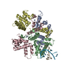
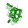
Proton-powered peptide transporter SbmA in lipid nanodisc
Map data
SbmA peptide transporter in nanodisc
Sample
Complex: Proton-driven peptide transporter SbmA in lipid nanodisc
Protein or peptide: SbmA
Function / homology
Function and homology information
microcin transmembrane transporter activity / microcin B17 transport / microcin transport / secondary active transmembrane transporter activity / peptide transport / peptide transmembrane transporter activity / protein transport / response to antibiotic / protein homodimerization activity / ATP binding / plasma membrane Similarity search - Function
Poland, United Kingdom, Japan, United States, 7 items
Organization
Grant number
Country
Polish National Science Centre
2016/21/B/CC1/00274
Poland
Foundation for Polish Science
TEAM TECH CORE FACILITY/2017-4/6
Poland
Polish National Science Centre
2019/35/D/NZ1/01770
Poland
Foundation for Polish Science
TEAM/2016-3/23
Poland
Biotechnology and Biological Sciences Research Council (BBSRC)
BB/H01778X/1
United Kingdom
Japan Agency for Medical Research and Development (AMED)
20am0101079
Japan
National Institutes of Health/National Cancer Institute (NIH/NCI)
GM31030
United States
Citation
Journal: Sci Adv / Year: 2021 Title: Molecular mechanism of SbmA, a promiscuous transporter exploited by antimicrobial peptides. Authors: Dmitry Ghilarov / Satomi Inaba-Inoue / Piotr Stepien / Feng Qu / Elizabeth Michalczyk / Zuzanna Pakosz / Norimichi Nomura / Satoshi Ogasawara / Graham Charles Walker / Sylvie Rebuffat / So ...Authors: Dmitry Ghilarov / Satomi Inaba-Inoue / Piotr Stepien / Feng Qu / Elizabeth Michalczyk / Zuzanna Pakosz / Norimichi Nomura / Satoshi Ogasawara / Graham Charles Walker / Sylvie Rebuffat / So Iwata / Jonathan Gardiner Heddle / Konstantinos Beis / Abstract: Antibiotic metabolites and antimicrobial peptides mediate competition between bacterial species. Many of them hijack inner and outer membrane proteins to enter cells. Sensitivity of enteric bacteria ...Antibiotic metabolites and antimicrobial peptides mediate competition between bacterial species. Many of them hijack inner and outer membrane proteins to enter cells. Sensitivity of enteric bacteria to multiple peptide antibiotics is controlled by the single inner membrane protein SbmA. To establish the molecular mechanism of peptide transport by SbmA and related BacA, we determined their cryo–electron microscopy structures at 3.2 and 6 Å local resolution, respectively. The structures show a previously unknown fold, defining a new class of secondary transporters named SbmA-like peptide transporters. The core domain includes conserved glutamates, which provide a pathway for proton translocation, powering transport. The structures show an outward-open conformation with a large cavity that can accommodate diverse substrates. We propose a molecular mechanism for antibacterial peptide uptake paving the way for creation of narrow-targeted therapeutics.
History
Deposition
Jul 6, 2021
-
Header (metadata) release
Sep 15, 2021
-
Map release
Sep 15, 2021
-
Update
Jan 19, 2022
-
Current status
Jan 19, 2022
Processing site: PDBe / Status: Released
-
Structure visualization
Movie
Surface view with section colored by density value
EMPIAR-10763 (Title: Proton-powered peptide transporter SbmA in lipid nanodisc Data size: 2.7 TB Data #1: Unaligned multi-frame movies of SbmA in lipid nanodisc [micrographs - multiframe])
In the structure databanks used in Yorodumi, some data are registered as the other names, "COVID-19 virus" and "2019-nCoV". Here are the details of the virus and the list of structure data.
Jan 31, 2019. EMDB accession codes are about to change! (news from PDBe EMDB page)
EMDB accession codes are about to change! (news from PDBe EMDB page)
The allocation of 4 digits for EMDB accession codes will soon come to an end. Whilst these codes will remain in use, new EMDB accession codes will include an additional digit and will expand incrementally as the available range of codes is exhausted. The current 4-digit format prefixed with “EMD-” (i.e. EMD-XXXX) will advance to a 5-digit format (i.e. EMD-XXXXX), and so on. It is currently estimated that the 4-digit codes will be depleted around Spring 2019, at which point the 5-digit format will come into force.
The EM Navigator/Yorodumi systems omit the EMD- prefix.
Related info.:Q: What is EMD? / ID/Accession-code notation in Yorodumi/EM Navigator
Yorodumi is a browser for structure data from EMDB, PDB, SASBDB, etc.
This page is also the successor to EM Navigator detail page, and also detail information page/front-end page for Omokage search.
The word "yorodu" (or yorozu) is an old Japanese word meaning "ten thousand". "mi" (miru) is to see.
Related info.:EMDB / PDB / SASBDB / Comparison of 3 databanks / Yorodumi Search / Aug 31, 2016. New EM Navigator & Yorodumi / Yorodumi Papers / Jmol/JSmol / Function and homology information / Changes in new EM Navigator and Yorodumi
 Movie
Movie Controller


 Open data
 Basic information
 Map data
 Sample
 Function and homology information
 Authors
 Poland,
  United Kingdom,
  Japan,
  United States, 7 items
  Citation
 Structure visualization
 Downloads & links
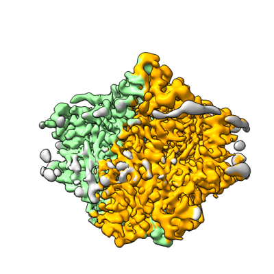 emd_13173.png
 http://ftp.pdbj.org/pub/emdb/structures/EMD-13173


 Z (Sec.)
 Y (Row.)
 X (Col.)


 Sample components
 Processing
 Electron microscopy
 FIELD EMISSION GUN

