 Movie
Movie Controller

[English] 日本語
 Yorodumi
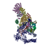
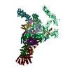
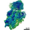
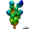
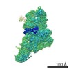
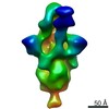
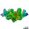
- PDB-7oqc: The U1 part of Saccharomyces cerevisiae spliceosomal pre-A comple... -
+ Open data

- Basic information
| Entry | Database: PDB / ID: 7oqc | ||||||
|---|---|---|---|---|---|---|---|
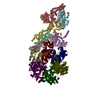
| Title | The U1 part of Saccharomyces cerevisiae spliceosomal pre-A complex (delta BS-A ACT1) | ||||||
 Components Components |
| ||||||
 Keywords Keywords | SPLICING / S. cerevisiae / pre-A complex / Prp5 / U1 snRNP / U2 snRNP / prespliceosome | ||||||
| Function / homology |  Function and homology information Function and homology informationmRNA splice site recognition / U4/U6 snRNP / 7-methylguanosine cap hypermethylation / pICln-Sm protein complex / positive regulation of mRNA splicing, via spliceosome / small nuclear ribonucleoprotein complex / SMN-Sm protein complex / spliceosomal tri-snRNP complex / splicing factor binding / snRNP binding ...mRNA splice site recognition / U4/U6 snRNP / 7-methylguanosine cap hypermethylation / pICln-Sm protein complex / positive regulation of mRNA splicing, via spliceosome / small nuclear ribonucleoprotein complex / SMN-Sm protein complex / spliceosomal tri-snRNP complex / splicing factor binding / snRNP binding / commitment complex / U2-type prespliceosome assembly / U1 snRNP / U2 snRNP / U4 snRNP / pre-mRNA 5'-splice site binding / U2-type prespliceosome / poly(U) RNA binding / precatalytic spliceosome / mRNA 5'-splice site recognition / spliceosomal complex assembly / Prp19 complex / U5 snRNP / spliceosomal snRNP assembly / U1 snRNA binding / U4/U6 x U5 tri-snRNP complex / catalytic step 2 spliceosome / spliceosomal complex / mRNA splicing, via spliceosome / mRNA binding / RNA binding / zinc ion binding / nucleus / cytoplasm / cytosol Similarity search - Function | ||||||
| Biological species |  | ||||||
| Method | ELECTRON MICROSCOPY / single particle reconstruction / cryo EM / Resolution: 4.1 Å | ||||||
 Authors Authors | Zhang, Z. / Rigo, N. / Dybkov, O. / Fourmann, J. / Will, C.L. / Kumar, V. / Urlaub, H. / Stark, H. / Luehrmann, R. | ||||||
| Funding support |  Germany, 1items Germany, 1items
| ||||||
 Citation Citation | Journal: Nature / Year: 2021 Title: Structural insights into how Prp5 proofreads the pre-mRNA branch site. Authors: Zhenwei Zhang / Norbert Rigo / Olexandr Dybkov / Jean-Baptiste Fourmann / Cindy L Will / Vinay Kumar / Henning Urlaub / Holger Stark / Reinhard Lührmann / Abstract: During the splicing of introns from precursor messenger RNAs (pre-mRNAs), the U2 small nuclear ribonucleoprotein (snRNP) must undergo stable integration into the spliceosomal A complex-a poorly ...During the splicing of introns from precursor messenger RNAs (pre-mRNAs), the U2 small nuclear ribonucleoprotein (snRNP) must undergo stable integration into the spliceosomal A complex-a poorly understood, multistep process that is facilitated by the DEAD-box helicase Prp5 (refs. ). During this process, the U2 small nuclear RNA (snRNA) forms an RNA duplex with the pre-mRNA branch site (the U2-BS helix), which is proofread by Prp5 at this stage through an unclear mechanism. Here, by deleting the branch-site adenosine (BS-A) or mutating the branch-site sequence of an actin pre-mRNA, we stall the assembly of spliceosomes in extracts from the yeast Saccharomyces cerevisiae directly before the A complex is formed. We then determine the three-dimensional structure of this newly identified assembly intermediate by cryo-electron microscopy. Our structure indicates that the U2-BS helix has formed in this pre-A complex, but is not yet clamped by the HEAT domain of the Hsh155 protein (Hsh155), which exhibits an open conformation. The structure further reveals a large-scale remodelling/repositioning of the U1 and U2 snRNPs during the formation of the A complex that is required to allow subsequent binding of the U4/U6.U5 tri-snRNP, but that this repositioning is blocked in the pre-A complex by the presence of Prp5. Our data suggest that binding of Hsh155 to the bulged BS-A of the U2-BS helix triggers closure of Hsh155, which in turn destabilizes Prp5 binding. Thus, Prp5 proofreads the branch site indirectly, hindering spliceosome assembly if branch-site mutations prevent the remodelling of Hsh155. Our data provide structural insights into how a spliceosomal helicase enhances the fidelity of pre-mRNA splicing. | ||||||
| History |
|
- Structure visualization
| Movie |
Movie viewer |
|---|---|
| Structure viewer | Molecule: MolmilJmol/JSmol |
- Downloads & links
-Download
| PDBx/mmCIF format | 7oqc.cif.gz | 846.5 KB | Display | PDBx/mmCIF format |
|---|---|---|---|---|
| PDB format | pdb7oqc.ent.gz | 636.4 KB | Display | PDB format |
| PDBx/mmJSON format | 7oqc.json.gz | Tree view | PDBx/mmJSON format | |
| Others |  Other downloads Other downloads |
-Validation report
| Arichive directory | https://data.pdbj.org/pub/pdb/validation_reports/oq/7oqcftp://data.pdbj.org/pub/pdb/validation_reports/oq/7oqc | HTTPS FTP |
|---|
-Related structure data
| Related structure data |  13029MC 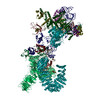 7oqbC 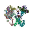 7oqeC M: map data used to model this data C: citing same article ( |
|---|---|
| Similar structure data |


-Links
 PDBj
PDBj


- Assembly
| Deposited unit | 
|
|---|---|
| 1 |
|
-Components
-Protein , 5 types, 5 molecules FGbHD
| #1: Protein | Mass: 57010.840 Da / Num. of mol.: 1 / Source method: isolated from a natural source / Source: (natural) |
|---|---|
| #6: Protein | Mass: 56575.277 Da / Num. of mol.: 1 / Source method: isolated from a natural source / Source: (natural) |
| #9: Protein | Mass: 22426.990 Da / Num. of mol.: 1 / Source method: isolated from a natural source / Source: (natural) |
| #16: Protein | Mass: 30245.885 Da / Num. of mol.: 1 / Source method: isolated from a natural source / Source: (natural) |
| #17: Protein | Mass: 74834.742 Da / Num. of mol.: 1 / Source method: isolated from a natural source / Source: (natural) |
-RNA chain , 2 types, 2 molecules I1
| #2: RNA chain | Mass: 111601.883 Da / Num. of mol.: 1 / Source method: isolated from a natural source / Source: (natural) |
|---|---|
| #5: RNA chain | Mass: 182114.516 Da / Num. of mol.: 1 / Source method: isolated from a natural source / Source: (natural) |
-U1 small nuclear ribonucleoprotein ... , 5 types, 5 molecules EJACB
| #3: Protein | Mass: 65222.020 Da / Num. of mol.: 1 / Source method: isolated from a natural source / Source: (natural) |
|---|---|
| #4: Protein | Mass: 71484.555 Da / Num. of mol.: 1 / Source method: isolated from a natural source / Source: (natural) |
| #7: Protein | Mass: 34438.105 Da / Num. of mol.: 1 / Source method: isolated from a natural source / Source: (natural) |
| #8: Protein | Mass: 27106.016 Da / Num. of mol.: 1 / Source method: isolated from a natural source / Source: (natural) |
| #18: Protein | Mass: 34506.148 Da / Num. of mol.: 1 / Source method: isolated from a natural source / Source: (natural) |
-Small nuclear ribonucleoprotein ... , 6 types, 6 molecules defghi
| #10: Protein | Mass: 11240.139 Da / Num. of mol.: 1 / Source method: isolated from a natural source / Source: (natural) |
|---|---|
| #11: Protein | Mass: 10385.098 Da / Num. of mol.: 1 / Source method: isolated from a natural source / Source: (natural) |
| #12: Protein | Mass: 9669.945 Da / Num. of mol.: 1 / Source method: isolated from a natural source / Source: (natural) |
| #13: Protein | Mass: 8490.809 Da / Num. of mol.: 1 / Source method: isolated from a natural source / Source: (natural) |
| #14: Protein | Mass: 16296.798 Da / Num. of mol.: 1 / Source method: isolated from a natural source / Source: (natural) |
| #15: Protein | Mass: 12876.066 Da / Num. of mol.: 1 / Source method: isolated from a natural source / Source: (natural) |
-Experimental details
-Experiment
| Experiment | Method: ELECTRON MICROSCOPY |
|---|---|
| EM experiment | Aggregation state: PARTICLE / 3D reconstruction method: single particle reconstruction |
- Sample preparation
| Component | Name: S. cerevisiae spliceosomal pre-A complex / Type: COMPLEX / Entity ID: all / Source: NATURAL |
|---|---|
| Molecular weight | Experimental value: NO |
| Source (natural) | Organism: |
| Buffer solution | pH: 7.9 |
| Specimen | Embedding applied: NO / Shadowing applied: NO / Staining applied: NO / Vitrification applied: YES |
| Specimen support | Grid material: COPPER / Grid type: Quantifoil R3.5/1 |
| Vitrification | Instrument: FEI VITROBOT MARK I / Cryogen name: ETHANE / Humidity: 100 % / Chamber temperature: 277 K |
- Electron microscopy imaging
| Experimental equipment |  Model: Titan Krios / Image courtesy: FEI Company |
|---|---|
| Microscopy | Model: FEI TITAN KRIOS |
| Electron gun | Electron source:  FIELD EMISSION GUN / Accelerating voltage: 300 kV / Illumination mode: SPOT SCAN FIELD EMISSION GUN / Accelerating voltage: 300 kV / Illumination mode: SPOT SCAN |
| Electron lens | Mode: BRIGHT FIELD / Cs: 0.01 mm / C2 aperture diameter: 100 µm / Alignment procedure: ZEMLIN TABLEAU |
| Specimen holder | Cryogen: NITROGEN / Specimen holder model: FEI TITAN KRIOS AUTOGRID HOLDER |
| Image recording | Average exposure time: 1.02 sec. / Electron dose: 44 e/Å2 / Detector mode: INTEGRATING / Film or detector model: FEI FALCON III (4k x 4k) |
- Processing
| EM software |
| ||||||||||||||||
|---|---|---|---|---|---|---|---|---|---|---|---|---|---|---|---|---|---|
| CTF correction | Type: PHASE FLIPPING AND AMPLITUDE CORRECTION | ||||||||||||||||
| 3D reconstruction | Resolution: 4.1 Å / Resolution method: FSC 0.143 CUT-OFF / Num. of particles: 217460 / Algorithm: FOURIER SPACE / Symmetry type: POINT |
