- EMDB-13031: The U1 part of Saccharomyces cerevisiae spliceosomal pre-A comple... -
+
Open data
ID or keywords:
Loading...
-
Basic information
Entry
Database: EMDB / ID: EMD-13031
Title
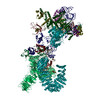
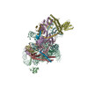
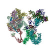
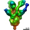
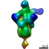
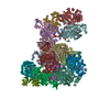
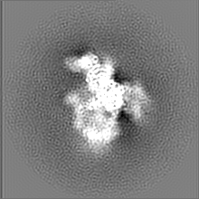
The U1 part of Saccharomyces cerevisiae spliceosomal pre-A complex (U257A)
Map data
U1 part of U257A pre-A complex
Sample
Complex: S. cerevisiae spliceosomal pre-A complex
Function / homology
Function and homology information
mRNA splice site recognition / U4/U6 snRNP / 7-methylguanosine cap hypermethylation / pICln-Sm protein complex / positive regulation of mRNA splicing, via spliceosome / small nuclear ribonucleoprotein complex / SMN-Sm protein complex / spliceosomal tri-snRNP complex / splicing factor binding / snRNP binding ...mRNA splice site recognition / U4/U6 snRNP / 7-methylguanosine cap hypermethylation / pICln-Sm protein complex / positive regulation of mRNA splicing, via spliceosome / small nuclear ribonucleoprotein complex / SMN-Sm protein complex / spliceosomal tri-snRNP complex / splicing factor binding / snRNP binding / commitment complex / U2-type prespliceosome assembly / U1 snRNP / U2 snRNP / U4 snRNP / pre-mRNA 5'-splice site binding / U2-type prespliceosome / poly(U) RNA binding / precatalytic spliceosome / mRNA 5'-splice site recognition / spliceosomal complex assembly / Prp19 complex / U5 snRNP / spliceosomal snRNP assembly / U1 snRNA binding / U4/U6 x U5 tri-snRNP complex / catalytic step 2 spliceosome / spliceosomal complex / mRNA splicing, via spliceosome / mRNA binding / RNA binding / zinc ion binding / nucleus / cytoplasm / cytosol Similarity search - Function
Luc7-related / LUC7 N_terminus / Snu56-like U1 small nuclear ribonucleoprotein component / : / : / Snu56-like U1 small nuclear ribonucleoprotein component / SNU71 RNA binding domain / SNU71 N-terminal / U1 small nuclear ribonucleoprotein C / : ...Luc7-related / LUC7 N_terminus / Snu56-like U1 small nuclear ribonucleoprotein component / : / : / Snu56-like U1 small nuclear ribonucleoprotein component / SNU71 RNA binding domain / SNU71 N-terminal / U1 small nuclear ribonucleoprotein C / : / PRP39 C-terminal HAT repeat / U1 small nuclear ribonucleoprotein of 70kDa N-terminal / : / U1 small nuclear ribonucleoprotein of 70kDa MW N terminal / : / U1-C, C2H2-type zinc finger / U1 zinc finger / PWI domain / PWI, domain in splicing factors / : / PRP39 N-terminal HAT repeat / Matrin/U1-C, C2H2-type zinc finger / Zinc finger matrin-type profile. / Small nuclear ribonucleoprotein Sm D2 / Small nuclear ribonucleoprotein Sm D3 / Small nuclear ribonucleoprotein E / Small nuclear ribonucleoprotein G / Small nuclear ribonucleoprotein F / Matrin/U1-C-like, C2H2-type zinc finger / U1-like zinc finger / Sm-like protein Lsm7/SmG / Like-Sm (LSM) domain containing protein, LSm4/SmD1/SmD3 / Sm-like protein Lsm6/SmF / LSM domain / LSM domain, eukaryotic/archaea-type / snRNP Sm proteins / HAT (Half-A-TPR) repeat / HAT (Half-A-TPR) repeats / : / Sm domain profile. / LSM domain superfamily / Zinc finger C2H2 superfamily / RNA recognition motif / RNA recognition motif / Eukaryotic RNA Recognition Motif (RRM) profile. / RNA recognition motif domain / RNA-binding domain superfamily / Tetratricopeptide-like helical domain superfamily / Nucleotide-binding alpha-beta plait domain superfamily Similarity search - Domain/homology
U1 small nuclear ribonucleoprotein A / Pre-mRNA-processing factor 39 / Small nuclear ribonucleoprotein-associated protein B / Small nuclear ribonucleoprotein G / Small nuclear ribonucleoprotein Sm D3 / U1 small nuclear ribonucleoprotein component SNU71 / Small nuclear ribonucleoprotein F / Protein NAM8 / U1 small nuclear ribonucleoprotein 70 kDa homolog / Small nuclear ribonucleoprotein Sm D1 ...U1 small nuclear ribonucleoprotein A / Pre-mRNA-processing factor 39 / Small nuclear ribonucleoprotein-associated protein B / Small nuclear ribonucleoprotein G / Small nuclear ribonucleoprotein Sm D3 / U1 small nuclear ribonucleoprotein component SNU71 / Small nuclear ribonucleoprotein F / Protein NAM8 / U1 small nuclear ribonucleoprotein 70 kDa homolog / Small nuclear ribonucleoprotein Sm D1 / U1 small nuclear ribonucleoprotein component PRP42 / 56 kDa U1 small nuclear ribonucleoprotein component / U1 small nuclear ribonucleoprotein C / Small nuclear ribonucleoprotein Sm D2 / Protein LUC7 / Small nuclear ribonucleoprotein E Similarity search - Component
Biological species
Saccharomyces cerevisiae (brewer's yeast)
Method
single particle reconstruction / cryo EM / Resolution: 7.5 Å
Journal: Nature / Year: 2021 Title: Structural insights into how Prp5 proofreads the pre-mRNA branch site. Authors: Zhenwei Zhang / Norbert Rigo / Olexandr Dybkov / Jean-Baptiste Fourmann / Cindy L Will / Vinay Kumar / Henning Urlaub / Holger Stark / Reinhard Lührmann / Abstract: During the splicing of introns from precursor messenger RNAs (pre-mRNAs), the U2 small nuclear ribonucleoprotein (snRNP) must undergo stable integration into the spliceosomal A complex-a poorly ...During the splicing of introns from precursor messenger RNAs (pre-mRNAs), the U2 small nuclear ribonucleoprotein (snRNP) must undergo stable integration into the spliceosomal A complex-a poorly understood, multistep process that is facilitated by the DEAD-box helicase Prp5 (refs. ). During this process, the U2 small nuclear RNA (snRNA) forms an RNA duplex with the pre-mRNA branch site (the U2-BS helix), which is proofread by Prp5 at this stage through an unclear mechanism. Here, by deleting the branch-site adenosine (BS-A) or mutating the branch-site sequence of an actin pre-mRNA, we stall the assembly of spliceosomes in extracts from the yeast Saccharomyces cerevisiae directly before the A complex is formed. We then determine the three-dimensional structure of this newly identified assembly intermediate by cryo-electron microscopy. Our structure indicates that the U2-BS helix has formed in this pre-A complex, but is not yet clamped by the HEAT domain of the Hsh155 protein (Hsh155), which exhibits an open conformation. The structure further reveals a large-scale remodelling/repositioning of the U1 and U2 snRNPs during the formation of the A complex that is required to allow subsequent binding of the U4/U6.U5 tri-snRNP, but that this repositioning is blocked in the pre-A complex by the presence of Prp5. Our data suggest that binding of Hsh155 to the bulged BS-A of the U2-BS helix triggers closure of Hsh155, which in turn destabilizes Prp5 binding. Thus, Prp5 proofreads the branch site indirectly, hindering spliceosome assembly if branch-site mutations prevent the remodelling of Hsh155. Our data provide structural insights into how a spliceosomal helicase enhances the fidelity of pre-mRNA splicing.
History
Deposition
Jun 3, 2021
-
Header (metadata) release
Aug 11, 2021
-
Map release
Aug 11, 2021
-
Update
Sep 29, 2021
-
Current status
Sep 29, 2021
Processing site: PDBe / Status: Released
-
Structure visualization
Movie
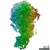
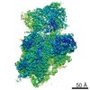
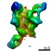
Surface view with section colored by density value
In the structure databanks used in Yorodumi, some data are registered as the other names, "COVID-19 virus" and "2019-nCoV". Here are the details of the virus and the list of structure data.
Jan 31, 2019. EMDB accession codes are about to change! (news from PDBe EMDB page)
EMDB accession codes are about to change! (news from PDBe EMDB page)
The allocation of 4 digits for EMDB accession codes will soon come to an end. Whilst these codes will remain in use, new EMDB accession codes will include an additional digit and will expand incrementally as the available range of codes is exhausted. The current 4-digit format prefixed with “EMD-” (i.e. EMD-XXXX) will advance to a 5-digit format (i.e. EMD-XXXXX), and so on. It is currently estimated that the 4-digit codes will be depleted around Spring 2019, at which point the 5-digit format will come into force.
The EM Navigator/Yorodumi systems omit the EMD- prefix.
Related info.:Q: What is EMD? / ID/Accession-code notation in Yorodumi/EM Navigator
Yorodumi is a browser for structure data from EMDB, PDB, SASBDB, etc.
This page is also the successor to EM Navigator detail page, and also detail information page/front-end page for Omokage search.
The word "yorodu" (or yorozu) is an old Japanese word meaning "ten thousand". "mi" (miru) is to see.
Related info.:EMDB / PDB / SASBDB / Comparison of 3 databanks / Yorodumi Search / Aug 31, 2016. New EM Navigator & Yorodumi / Yorodumi Papers / Jmol/JSmol / Function and homology information / Changes in new EM Navigator and Yorodumi
 Movie
Movie Controller

 Yorodumi
 Open data
 Basic information
 Map data
 Sample
 Function and homology information
 Authors
 Germany, 1 items
  Citation
 Structure visualization
 Downloads & links
 emd_13031.png
 http://ftp.pdbj.org/pub/emdb/structures/EMD-13031


 Z (Sec.)
 Y (Row.)
 X (Col.)


 Sample components
 Processing
 Electron microscopy
 FIELD EMISSION GUN
