 Movie
Movie Controller

[English] 日本語
 Yorodumi
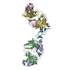
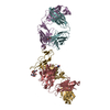
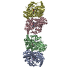
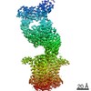
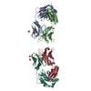
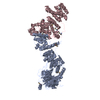
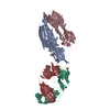
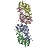
- PDB-7ob4: Cryo-EM structure of a twisted-dimer transthyretin amyloid fibril... -
+ Open data

- Basic information
| Entry | Database: PDB / ID: 7ob4 | ||||||
|---|---|---|---|---|---|---|---|
| Title | Cryo-EM structure of a twisted-dimer transthyretin amyloid fibril from vitreous body of the eye | ||||||
 Components Components | Transthyretin | ||||||
 Keywords Keywords | PROTEIN FIBRIL / AMYLOID FIBRIL / TRANSTHYRETIN / MISFOLDING / ATTR AMYLOIDOSIS / V30M VARIANT / EX VIVO / VITREOUS BODY | ||||||
| Function / homology |  Function and homology information Function and homology informationDefective visual phototransduction due to STRA6 loss of function / negative regulation of glomerular filtration / The canonical retinoid cycle in rods (twilight vision) / purine nucleobase metabolic process / hormone binding / Non-integrin membrane-ECM interactions / phototransduction, visible light / molecular sequestering activity / retinoid metabolic process / Retinoid metabolism and transport ...Defective visual phototransduction due to STRA6 loss of function / negative regulation of glomerular filtration / The canonical retinoid cycle in rods (twilight vision) / purine nucleobase metabolic process / hormone binding / Non-integrin membrane-ECM interactions / phototransduction, visible light / molecular sequestering activity / retinoid metabolic process / Retinoid metabolism and transport / hormone activity / azurophil granule lumen / Amyloid fiber formation / Neutrophil degranulation / protein-containing complex binding / protein-containing complex / : / extracellular exosome / extracellular region / identical protein binding Similarity search - Function | ||||||
| Biological species |  Homo sapiens (human) Homo sapiens (human) | ||||||
| Method | ELECTRON MICROSCOPY / helical reconstruction / cryo EM / Resolution: 3.22 Å | ||||||
 Authors Authors | Iakovleva, I. / Sauer-Eriksson, A.E. | ||||||
| Funding support |  Sweden, 1items Sweden, 1items
| ||||||
 Citation Citation | Journal: Nat Commun / Year: 2021 Title: Structural basis for transthyretin amyloid formation in vitreous body of the eye. Authors: Irina Iakovleva / Michael Hall / Melanie Oelker / Linda Sandblad / Intissar Anan / A Elisabeth Sauer-Eriksson / Abstract: Amyloid transthyretin (ATTR) amyloidosis is characterized by the abnormal accumulation of ATTR fibrils in multiple organs. However, the structure of ATTR fibrils from the eye is poorly understood. ...Amyloid transthyretin (ATTR) amyloidosis is characterized by the abnormal accumulation of ATTR fibrils in multiple organs. However, the structure of ATTR fibrils from the eye is poorly understood. Here, we used cryo-EM to structurally characterize vitreous body ATTR fibrils. These structures were distinct from previously characterized heart fibrils, even though both have the same mutation and type A pathology. Differences were observed at several structural levels: in both the number and arrangement of protofilaments, and the conformation of the protein fibril in each layer of protofilaments. Thus, our results show that ATTR protein structure and its assembly into protofilaments in the type A fibrils can vary between patients carrying the same mutation. By analyzing and matching the interfaces between the amino acids in the ATTR fibril with those in the natively folded TTR, we are able to propose a mechanism for the structural conversion of TTR into a fibrillar form. | ||||||
| History |
|
- Structure visualization
| Movie |
Movie viewer |
|---|---|
| Structure viewer | Molecule: MolmilJmol/JSmol |
- Downloads & links
-Download
| PDBx/mmCIF format | 7ob4.cif.gz | 226.3 KB | Display | PDBx/mmCIF format |
|---|---|---|---|---|
| PDB format | pdb7ob4.ent.gz | 182.7 KB | Display | PDB format |
| PDBx/mmJSON format | 7ob4.json.gz | Tree view | PDBx/mmJSON format | |
| Others |  Other downloads Other downloads |
-Validation report
| Arichive directory | https://data.pdbj.org/pub/pdb/validation_reports/ob/7ob4ftp://data.pdbj.org/pub/pdb/validation_reports/ob/7ob4 | HTTPS FTP |
|---|
-Related structure data
| Related structure data |  12794MC M: map data used to model this data C: citing same article ( |
|---|---|
| Similar structure data |
-Links
 PDBj
PDBj


- Assembly
| Deposited unit | 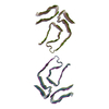
|
|---|---|
| 1 |
|
-Components
| #1: Protein | Mass: 13809.426 Da / Num. of mol.: 14 / Source method: isolated from a natural source / Source: (natural) Homo sapiens (human) / References: UniProt: P02766 |
|---|
-Experimental details
-Experiment
| Experiment | Method: ELECTRON MICROSCOPY |
|---|---|
| EM experiment | Aggregation state: HELICAL ARRAY / 3D reconstruction method: helical reconstruction |
- Sample preparation
| Component | Name: Transthyretin derived amyloid fibril (V30M) from vitreous body Type: TISSUE / Entity ID: all / Source: NATURAL |
|---|---|
| Source (natural) | Organism: Homo sapiens (human) / Organ: Vitreous body of the eye |
| Buffer solution | pH: 7.4 |
| Specimen | Embedding applied: NO / Shadowing applied: NO / Staining applied: NO / Vitrification applied: YES |
| Vitrification | Instrument: FEI VITROBOT MARK IV / Cryogen name: ETHANE |
- Electron microscopy imaging
| Experimental equipment |  Model: Titan Krios / Image courtesy: FEI Company |
|---|---|
| Microscopy | Model: FEI TITAN KRIOS |
| Electron gun | Electron source:  FIELD EMISSION GUN / Accelerating voltage: 300 kV / Illumination mode: FLOOD BEAM FIELD EMISSION GUN / Accelerating voltage: 300 kV / Illumination mode: FLOOD BEAM |
| Electron lens | Mode: BRIGHT FIELD |
| Image recording | Electron dose: 28.4 e/Å2 / Film or detector model: GATAN K2 SUMMIT (4k x 4k) |
- Processing
| EM software |
| ||||||||||||||||||||||||||||
|---|---|---|---|---|---|---|---|---|---|---|---|---|---|---|---|---|---|---|---|---|---|---|---|---|---|---|---|---|---|
| CTF correction | Type: PHASE FLIPPING AND AMPLITUDE CORRECTION | ||||||||||||||||||||||||||||
| Helical symmerty | Angular rotation/subunit: -0.552 ° / Axial rise/subunit: 4.72 Å / Axial symmetry: C2 | ||||||||||||||||||||||||||||
| Particle selection | Num. of particles selected: 130212 | ||||||||||||||||||||||||||||
| 3D reconstruction | Resolution: 3.22 Å / Resolution method: FSC 0.143 CUT-OFF / Num. of particles: 27778 / Symmetry type: HELICAL | ||||||||||||||||||||||||||||
| Atomic model building | Protocol: RIGID BODY FIT / Space: REAL | ||||||||||||||||||||||||||||
| Atomic model building | PDB-ID: 6SDZ Accession code: 6SDZ / Source name: PDB / Type: experimental model |

