 Movie
Movie Controller

[English] 日本語
 Yorodumi
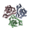
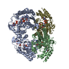
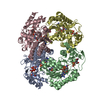
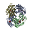
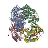
- PDB-7ngh: Structure of glutamate transporter homologue in complex with Sybody -
+ Open data

- Basic information
| Entry | Database: PDB / ID: 7ngh | |||||||||||||||||||||
|---|---|---|---|---|---|---|---|---|---|---|---|---|---|---|---|---|---|---|---|---|---|---|
| Title | Structure of glutamate transporter homologue in complex with Sybody | |||||||||||||||||||||
 Components Components |
| |||||||||||||||||||||
 Keywords Keywords | TRANSPORT PROTEIN / glutamate transporter homologue / GltTk / amino acid transport / membrane protein | |||||||||||||||||||||
| Function / homology |  Function and homology information Function and homology informationdicarboxylic acid transport / symporter activity / membrane / plasma membrane Similarity search - Function | |||||||||||||||||||||
| Biological species |   Thermococcus kodakarensis KOD1 (archaea) Thermococcus kodakarensis KOD1 (archaea)synthetic construct (others) | |||||||||||||||||||||
| Method | ELECTRON MICROSCOPY / single particle reconstruction / cryo EM / Resolution: 3.5 Å | |||||||||||||||||||||
 Authors Authors | Arkhipova, V. / Slotboom, D.J. / Guskov, A. | |||||||||||||||||||||
 Citation Citation | Journal: Commun Biol / Year: 2021 Title: Kinetic mechanism of Na-coupled aspartate transport catalyzed by Glt. Authors: Gianluca Trinco / Valentina Arkhipova / Alisa A Garaeva / Cedric A J Hutter / Markus A Seeger / Albert Guskov / Dirk J Slotboom /    Abstract: It is well-established that the secondary active transporters Glt and Glt catalyze coupled uptake of aspartate and three sodium ions, but insight in the kinetic mechanism of transport is fragmentary. ...It is well-established that the secondary active transporters Glt and Glt catalyze coupled uptake of aspartate and three sodium ions, but insight in the kinetic mechanism of transport is fragmentary. Here, we systematically measured aspartate uptake rates in proteoliposomes containing purified Glt, and derived the rate equation for a mechanism in which two sodium ions bind before and another after aspartate. Re-analysis of existing data on Glt using this equation allowed for determination of the turnover number (0.14 s), without the need for error-prone protein quantification. To overcome the complication that purified transporters may adopt right-side-out or inside-out membrane orientations upon reconstitution, thereby confounding the kinetic analysis, we employed a rapid method using synthetic nanobodies to inactivate one population. Oppositely oriented Glt proteins showed the same transport kinetics, consistent with the use of an identical gating element on both sides of the membrane. Our work underlines the value of bona fide transport experiments to reveal mechanistic features of Na-aspartate symport that cannot be observed in detergent solution. Combined with previous pre-equilibrium binding studies, a full kinetic mechanism of structurally characterized aspartate transporters of the SLC1A family is now emerging. | |||||||||||||||||||||
| History |
|
- Structure visualization
| Movie |
Movie viewer |
|---|---|
| Structure viewer | Molecule: MolmilJmol/JSmol |
- Downloads & links
-Download
| PDBx/mmCIF format | 7ngh.cif.gz | 230.5 KB | Display | PDBx/mmCIF format |
|---|---|---|---|---|
| PDB format | pdb7ngh.ent.gz | 186.9 KB | Display | PDB format |
| PDBx/mmJSON format | 7ngh.json.gz | Tree view | PDBx/mmJSON format | |
| Others |  Other downloads Other downloads |
-Validation report
| Arichive directory | https://data.pdbj.org/pub/pdb/validation_reports/ng/7nghftp://data.pdbj.org/pub/pdb/validation_reports/ng/7ngh | HTTPS FTP |
|---|
-Related structure data
| Related structure data | 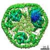 12314MC M: map data used to model this data C: citing same article ( |
|---|---|
| Similar structure data |


-Links
 PDBj
PDBj


- Assembly
| Deposited unit | 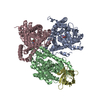
|
|---|---|
| 1 |
|
-Components
| #1: Protein | Mass: 46409.070 Da / Num. of mol.: 3 Source method: isolated from a genetically manipulated source Source: (gene. exp.) Thermococcus kodakarensis KOD1 (archaea)Gene: TK0986 / Production host:  #2: Antibody | | Mass: 15862.396 Da / Num. of mol.: 1 Source method: isolated from a genetically manipulated source Details: Hexa His tag at C-terminus / Source: (gene. exp.) synthetic construct (others) / Production host: #3: Chemical |   Type: L-peptide linking / Mass: 133.103 Da / Num. of mol.: 3 / Source method: obtained synthetically / Formula: C4H7NO4 Type: L-peptide linking / Mass: 133.103 Da / Num. of mol.: 3 / Source method: obtained synthetically / Formula: C4H7NO4Has ligand of interest | N | Has protein modification | Y | |
|---|
-Experimental details
-Experiment
| Experiment | Method: ELECTRON MICROSCOPY |
|---|---|
| EM experiment | Aggregation state: PARTICLE / 3D reconstruction method: single particle reconstruction |
- Sample preparation
| Component |
| ||||||||||||||||||||||||
|---|---|---|---|---|---|---|---|---|---|---|---|---|---|---|---|---|---|---|---|---|---|---|---|---|---|
| Molecular weight | Experimental value: NO | ||||||||||||||||||||||||
| Source (natural) |
| ||||||||||||||||||||||||
| Buffer solution | pH: 8 | ||||||||||||||||||||||||
| Specimen | Conc.: 1 mg/ml / Embedding applied: NO / Shadowing applied: NO / Staining applied: NO / Vitrification applied: YES | ||||||||||||||||||||||||
| Vitrification | Cryogen name: ETHANE |
- Electron microscopy imaging
| Experimental equipment |  Model: Talos Arctica / Image courtesy: FEI Company |
|---|---|
| Microscopy | Model: FEI TALOS ARCTICA |
| Electron gun | Electron source:  FIELD EMISSION GUN / Accelerating voltage: 200 kV / Illumination mode: FLOOD BEAM FIELD EMISSION GUN / Accelerating voltage: 200 kV / Illumination mode: FLOOD BEAM |
| Electron lens | Mode: BRIGHT FIELD |
| Image recording | Electron dose: 53 e/Å2 / Film or detector model: GATAN K2 QUANTUM (4k x 4k) |
- Processing
| Software | Name: PHENIX / Version: 1.16_3549: / Classification: refinement | ||||||||||||||||||||||||
|---|---|---|---|---|---|---|---|---|---|---|---|---|---|---|---|---|---|---|---|---|---|---|---|---|---|
| EM software | Name: PHENIX / Category: model refinement | ||||||||||||||||||||||||
| CTF correction | Type: NONE | ||||||||||||||||||||||||
| 3D reconstruction | Resolution: 3.5 Å / Resolution method: FSC 0.143 CUT-OFF / Num. of particles: 53983 / Symmetry type: POINT | ||||||||||||||||||||||||
| Refine LS restraints |
|
