[English] 日本語
 Yorodumi
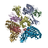
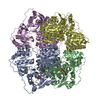
- EMDB-12314: Structure of glutamate transporter homologue in complex with Sybody -
+ Open data
- Basic information
| Entry | Database: EMDB / ID: EMD-12314 | |||||||||
|---|---|---|---|---|---|---|---|---|---|---|
| Title | Structure of glutamate transporter homologue in complex with Sybody | |||||||||
 Map data Map data | ||||||||||
 Sample Sample |
| |||||||||
 Keywords Keywords | glutamate transporter homologue / GltTk / amino acid transport / membrane protein / TRANSPORT PROTEIN | |||||||||
| Function / homology |  Function and homology information Function and homology informationdicarboxylic acid transport / symporter activity / membrane / plasma membrane Similarity search - Function | |||||||||
| Biological species |   Thermococcus kodakarensis KOD1 (archaea) / synthetic construct (others) Thermococcus kodakarensis KOD1 (archaea) / synthetic construct (others) | |||||||||
| Method | single particle reconstruction / cryo EM / Resolution: 3.5 Å | |||||||||
 Authors Authors | Arkhipova V / Slotboom DJ | |||||||||
 Citation Citation |  Journal: Commun Biol / Year: 2021 Journal: Commun Biol / Year: 2021Title: Kinetic mechanism of Na-coupled aspartate transport catalyzed by Glt. Authors: Gianluca Trinco / Valentina Arkhipova / Alisa A Garaeva / Cedric A J Hutter / Markus A Seeger / Albert Guskov / Dirk J Slotboom /    Abstract: It is well-established that the secondary active transporters Glt and Glt catalyze coupled uptake of aspartate and three sodium ions, but insight in the kinetic mechanism of transport is fragmentary. ...It is well-established that the secondary active transporters Glt and Glt catalyze coupled uptake of aspartate and three sodium ions, but insight in the kinetic mechanism of transport is fragmentary. Here, we systematically measured aspartate uptake rates in proteoliposomes containing purified Glt, and derived the rate equation for a mechanism in which two sodium ions bind before and another after aspartate. Re-analysis of existing data on Glt using this equation allowed for determination of the turnover number (0.14 s), without the need for error-prone protein quantification. To overcome the complication that purified transporters may adopt right-side-out or inside-out membrane orientations upon reconstitution, thereby confounding the kinetic analysis, we employed a rapid method using synthetic nanobodies to inactivate one population. Oppositely oriented Glt proteins showed the same transport kinetics, consistent with the use of an identical gating element on both sides of the membrane. Our work underlines the value of bona fide transport experiments to reveal mechanistic features of Na-aspartate symport that cannot be observed in detergent solution. Combined with previous pre-equilibrium binding studies, a full kinetic mechanism of structurally characterized aspartate transporters of the SLC1A family is now emerging. | |||||||||
| History |
|
- Structure visualization
| Movie |
 Movie viewer Movie viewer |
|---|---|
| Structure viewer | EM map:  SurfView SurfView Molmil Molmil Jmol/JSmol Jmol/JSmol |
| Supplemental images |
- Downloads & links
-EMDB archive
| Map data |  emd_12314.map.gz emd_12314.map.gz | 32.1 MB |  EMDB map data format EMDB map data format | |
|---|---|---|---|---|
| Header (meta data) |  emd-12314-v30.xml emd-12314-v30.xml emd-12314.xml emd-12314.xml | 13.4 KB 13.4 KB | Display Display |  EMDB header EMDB header |
| Images |  emd_12314.png emd_12314.png | 51.6 KB | ||
| Filedesc metadata |  emd-12314.cif.gz emd-12314.cif.gz | 5.7 KB | ||
| Archive directory |  http://ftp.pdbj.org/pub/emdb/structures/EMD-12314 http://ftp.pdbj.org/pub/emdb/structures/EMD-12314 ftp://ftp.pdbj.org/pub/emdb/structures/EMD-12314 ftp://ftp.pdbj.org/pub/emdb/structures/EMD-12314 | HTTPS FTP |
-Related structure data
| Related structure data |  7nghMC M: atomic model generated by this map C: citing same article ( |
|---|---|
| Similar structure data |
- Links
Links
| EMDB pages |  EMDB (EBI/PDBe) / EMDB (EBI/PDBe) /  EMDataResource EMDataResource |
|---|---|
| Related items in Molecule of the Month |
- Map
Map
| File |  Download / File: emd_12314.map.gz / Format: CCP4 / Size: 64 MB / Type: IMAGE STORED AS FLOATING POINT NUMBER (4 BYTES) Download / File: emd_12314.map.gz / Format: CCP4 / Size: 64 MB / Type: IMAGE STORED AS FLOATING POINT NUMBER (4 BYTES) | ||||||||||||||||||||||||||||||||||||||||||||||||||||||||||||||||||||
|---|---|---|---|---|---|---|---|---|---|---|---|---|---|---|---|---|---|---|---|---|---|---|---|---|---|---|---|---|---|---|---|---|---|---|---|---|---|---|---|---|---|---|---|---|---|---|---|---|---|---|---|---|---|---|---|---|---|---|---|---|---|---|---|---|---|---|---|---|---|
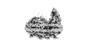
| Projections & slices | Image control
Images are generated by Spider. | ||||||||||||||||||||||||||||||||||||||||||||||||||||||||||||||||||||
| Voxel size | X=Y=Z: 1.012 Å | ||||||||||||||||||||||||||||||||||||||||||||||||||||||||||||||||||||
| Density |
| ||||||||||||||||||||||||||||||||||||||||||||||||||||||||||||||||||||
| Symmetry | Space group: 1 | ||||||||||||||||||||||||||||||||||||||||||||||||||||||||||||||||||||
| Details | EMDB XML:
CCP4 map header:
| ||||||||||||||||||||||||||||||||||||||||||||||||||||||||||||||||||||
-Supplemental data
- Sample components
-Entire : GltTk complex with sybody
| Entire | Name: GltTk complex with sybody |
|---|---|
| Components |
|
-Supramolecule #1: GltTk complex with sybody
| Supramolecule | Name: GltTk complex with sybody / type: complex / ID: 1 / Parent: 0 / Macromolecule list: #1-#2 |
|---|
-Supramolecule #2: Thermococcus kodakarensis KOD1
| Supramolecule | Name: Thermococcus kodakarensis KOD1 / type: complex / ID: 2 / Parent: 1 / Macromolecule list: #1 |
|---|---|
| Source (natural) | Organism:   Thermococcus kodakarensis KOD1 (archaea) Thermococcus kodakarensis KOD1 (archaea) |
-Supramolecule #3: synthetic construct
| Supramolecule | Name: synthetic construct / type: complex / ID: 3 / Parent: 1 / Macromolecule list: #2 |
|---|---|
| Source (natural) | Organism: synthetic construct (others) / Synthetically produced: Yes |
-Macromolecule #1: Proton/glutamate symporter, SDF family
| Macromolecule | Name: Proton/glutamate symporter, SDF family / type: protein_or_peptide / ID: 1 / Number of copies: 3 / Enantiomer: LEVO |
|---|---|
| Source (natural) | Organism:   Thermococcus kodakarensis KOD1 (archaea) Thermococcus kodakarensis KOD1 (archaea) |
| Molecular weight | Theoretical: 46.40907 KDa |
| Recombinant expression | Organism:  |
| Sequence | String: MGKSLLRRYL DYPVLWKILW GLVLGAVFGL IAGHFGYAGA VKTYIKPFGD LFVRLLKMLV MPIVLASLVV GAASISPARL GRVGVKIVV YYLATSAMAV FFGLIVGRLF NVGANVNLGS GTGKAIEAQP PSLVQTLLNI VPTNPFASLA KGEVLPVIFF A IILGIAIT ...String: MGKSLLRRYL DYPVLWKILW GLVLGAVFGL IAGHFGYAGA VKTYIKPFGD LFVRLLKMLV MPIVLASLVV GAASISPARL GRVGVKIVV YYLATSAMAV FFGLIVGRLF NVGANVNLGS GTGKAIEAQP PSLVQTLLNI VPTNPFASLA KGEVLPVIFF A IILGIAIT YLMNRNEERV RKSAETLLRV FDGLAEAMYL IVGGVMQYAP IGVFALIAYV MAEQGVRVVG PLAKVVGAVY TG LFLQIVI TYFILLKVFG IDPIKFIRKA KDAMITAFVT RSSSGTLPVT MRVAEEEMGV DKGIFSFTLP LGATINMDGT ALY QGVTVL FVANAIGHPL TLGQQLVVVL TAVLASIGTA GVPGAGAIML AMVLQSVGLD LTPGSPVALA YAMILGIDAI LDMG RTMVN VTGDLAGTVI VAKTEKELDE SKWISHHHHH H UniProtKB: Proton/glutamate symporter, SDF family |
-Macromolecule #2: Sybody 1
| Macromolecule | Name: Sybody 1 / type: protein_or_peptide / ID: 2 / Details: Hexa His tag at C-terminus / Number of copies: 1 / Enantiomer: LEVO |
|---|---|
| Source (natural) | Organism: synthetic construct (others) |
| Molecular weight | Theoretical: 15.862396 KDa |
| Recombinant expression | Organism:  |
| Sequence | String: GSSSQVQLVE SGGGLVQAGG SLRLSCAASG FPVDSQFMHW YRQAPGKERE WVAAIESYGD ETYYADSVKG RFTISRDNAK NTVYLQMNS LKPEDTAVYY CRVLVGWGYY GQGTQVTVSA GRAGEQKLIS EEDLNSAVDH HHHHH |
-Macromolecule #3: ASPARTIC ACID
| Macromolecule | Name: ASPARTIC ACID / type: ligand / ID: 3 / Number of copies: 3 / Formula: ASP |
|---|---|
| Molecular weight | Theoretical: 133.103 Da |
| Chemical component information | 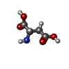 ChemComp-ASP: |
-Experimental details
-Structure determination
| Method | cryo EM |
|---|---|
 Processing Processing | single particle reconstruction |
| Aggregation state | particle |
- Sample preparation
| Concentration | 1 mg/mL |
|---|---|
| Buffer | pH: 8 |
| Vitrification | Cryogen name: ETHANE |
- Electron microscopy
| Microscope | FEI TALOS ARCTICA |
|---|---|
| Image recording | Film or detector model: GATAN K2 QUANTUM (4k x 4k) / Average electron dose: 53.0 e/Å2 |
| Electron beam | Acceleration voltage: 200 kV / Electron source:  FIELD EMISSION GUN FIELD EMISSION GUN |
| Electron optics | Illumination mode: FLOOD BEAM / Imaging mode: BRIGHT FIELD |
| Experimental equipment |  Model: Talos Arctica / Image courtesy: FEI Company |
 Movie
Movie Controller






 Z (Sec.)
 Y (Row.)
 X (Col.)


