 Movie
Movie Controller

[English] 日本語
 Yorodumi
- PDB-7n0l: Cryo-EM structure of TACAN in the H196A H197A mutant form (TMEM120A) -
+ Open data

- Basic information
| Entry | Database: PDB / ID: 7n0l | ||||||
|---|---|---|---|---|---|---|---|
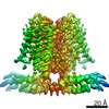
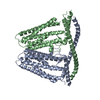
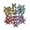
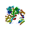
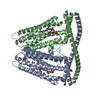
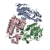
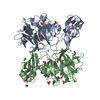
| Title | Cryo-EM structure of TACAN in the H196A H197A mutant form (TMEM120A) | ||||||
 Components Components | Ion channel TACAN | ||||||
 Keywords Keywords | MEMBRANE PROTEIN / lipid metabolism / coenzyme A | ||||||
| Function / homology |  Function and homology information Function and homology informationcoenzyme A binding / protein heterooligomerization / nuclear inner membrane / monoatomic ion channel activity / detection of mechanical stimulus involved in sensory perception of pain / fat cell differentiation / antiviral innate immune response / protein homooligomerization / monoatomic ion transmembrane transport / endoplasmic reticulum ...coenzyme A binding / protein heterooligomerization / nuclear inner membrane / monoatomic ion channel activity / detection of mechanical stimulus involved in sensory perception of pain / fat cell differentiation / antiviral innate immune response / protein homooligomerization / monoatomic ion transmembrane transport / endoplasmic reticulum / membrane / plasma membrane Similarity search - Function | ||||||
| Biological species |  | ||||||
| Method | ELECTRON MICROSCOPY / single particle reconstruction / cryo EM / Resolution: 2.8 Å | ||||||
 Authors Authors | Niu, Y. / Tao, X. / MacKinnon, R. | ||||||
| Funding support |  United States, 1items United States, 1items
| ||||||
 Citation Citation | Journal: Elife / Year: 2021 Title: Analysis of the mechanosensor channel functionality of TACAN. Authors: Yiming Niu / Xiao Tao / George Vaisey / Paul Dominic B Olinares / Hanan Alwaseem / Brian T Chait / Roderick MacKinnon / Abstract: Mechanosensitive ion channels mediate transmembrane ion currents activated by mechanical forces. A mechanosensitive ion channel called TACAN was recently reported. We began to study TACAN with the ...Mechanosensitive ion channels mediate transmembrane ion currents activated by mechanical forces. A mechanosensitive ion channel called TACAN was recently reported. We began to study TACAN with the intent to understand how it senses mechanical forces and functions as an ion channel. Using cellular patch-recording methods, we failed to identify mechanosensitive ion channel activity. Using membrane reconstitution methods, we found that TACAN, at high protein concentrations, produces heterogeneous conduction levels that are not mechanosensitive and are most consistent with disruptions of the lipid bilayer. We determined the structure of TACAN using single-particle cryo-electron microscopy and observed that it is a symmetrical dimeric transmembrane protein. Each protomer contains an intracellular-facing cleft with a coenzyme A cofactor, confirmed by mass spectrometry. The TACAN protomer is related in three-dimensional structure to a fatty acid elongase, ELOVL7. Whilst its physiological function remains unclear, we anticipate that TACAN is not a mechanosensitive ion channel. | ||||||
| History |
|
- Structure visualization
| Movie |
Movie viewer |
|---|---|
| Structure viewer | Molecule: MolmilJmol/JSmol |
- Downloads & links
-Download
| PDBx/mmCIF format | 7n0l.cif.gz | 124.9 KB | Display | PDBx/mmCIF format |
|---|---|---|---|---|
| PDB format | pdb7n0l.ent.gz | 97 KB | Display | PDB format |
| PDBx/mmJSON format | 7n0l.json.gz | Tree view | PDBx/mmJSON format | |
| Others |  Other downloads Other downloads |
-Validation report
| Arichive directory | https://data.pdbj.org/pub/pdb/validation_reports/n0/7n0lftp://data.pdbj.org/pub/pdb/validation_reports/n0/7n0l | HTTPS FTP |
|---|
-Related structure data
| Related structure data | 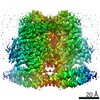 24108MC 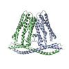 7n0kC C: citing same article ( M: map data used to model this data |
|---|---|
| Similar structure data |


-Links
 PDBj
PDBj


- Assembly
| Deposited unit | 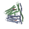
|
|---|---|
| 1 |
|
-Components
| #1: Protein | Mass: 40663.160 Da / Num. of mol.: 2 / Mutation: H196A, H197A Source method: isolated from a genetically manipulated source Source: (gene. exp.)  Homo sapiens (human) / References: UniProt: Q8C1E7 Homo sapiens (human) / References: UniProt: Q8C1E7#2: Chemical |   Mass: 767.534 Da / Num. of mol.: 2 / Source method: obtained synthetically / Formula: C21H36N7O16P3S / Feature type: SUBJECT OF INVESTIGATION Mass: 767.534 Da / Num. of mol.: 2 / Source method: obtained synthetically / Formula: C21H36N7O16P3S / Feature type: SUBJECT OF INVESTIGATIONHas ligand of interest | Y | |
|---|
-Experimental details
-Experiment
| Experiment | Method: ELECTRON MICROSCOPY |
|---|---|
| EM experiment | Aggregation state: PARTICLE / 3D reconstruction method: single particle reconstruction |
- Sample preparation
| Component | Name: Homo-dimeric assembly of H196A H197A mutant TACAN(TMEM120A) Type: COMPLEX / Entity ID: #1 / Source: RECOMBINANT |
|---|---|
| Molecular weight | Units: KILODALTONS/NANOMETER / Experimental value: NO |
| Source (natural) | Organism: |
| Source (recombinant) | Organism: Homo sapiens (human) |
| Buffer solution | pH: 7.4 Details: 20 mM HEPES pH 7.4, 250 mM NaCl, and 0.06% Digitonin (w/v) |
| Specimen | Conc.: 6 mg/ml / Embedding applied: NO / Shadowing applied: NO / Staining applied: NO / Vitrification applied: YES |
| Specimen support | Grid material: GOLD / Grid mesh size: 400 divisions/in. / Grid type: Quantifoil R1.2/1.3 |
| Vitrification | Instrument: FEI VITROBOT MARK IV / Cryogen name: ETHANE / Humidity: 95 % / Chamber temperature: 298 K |
- Electron microscopy imaging
| Experimental equipment |  Model: Titan Krios / Image courtesy: FEI Company |
|---|---|
| Microscopy | Model: FEI TITAN KRIOS |
| Electron gun | Electron source:  FIELD EMISSION GUN / Accelerating voltage: 300 kV / Illumination mode: FLOOD BEAM FIELD EMISSION GUN / Accelerating voltage: 300 kV / Illumination mode: FLOOD BEAM |
| Electron lens | Mode: DARK FIELD / C2 aperture diameter: 100 µm |
| Specimen holder | Cryogen: NITROGEN / Specimen holder model: FEI TITAN KRIOS AUTOGRID HOLDER |
| Image recording | Average exposure time: 1.5 sec. / Electron dose: 56.6 e/Å2 / Film or detector model: GATAN K3 (6k x 4k) |
- Processing
| Software | Name: PHENIX / Version: dev_3751: / Classification: refinement | ||||||||||||||||||||||||||||||||||||||||||||||||||||
|---|---|---|---|---|---|---|---|---|---|---|---|---|---|---|---|---|---|---|---|---|---|---|---|---|---|---|---|---|---|---|---|---|---|---|---|---|---|---|---|---|---|---|---|---|---|---|---|---|---|---|---|---|---|
| EM software |
| ||||||||||||||||||||||||||||||||||||||||||||||||||||
| CTF correction | Type: NONE | ||||||||||||||||||||||||||||||||||||||||||||||||||||
| Symmetry | Point symmetry: C2 (2 fold cyclic) | ||||||||||||||||||||||||||||||||||||||||||||||||||||
| 3D reconstruction | Resolution: 2.8 Å / Resolution method: FSC 0.143 CUT-OFF / Num. of particles: 155946 / Symmetry type: POINT | ||||||||||||||||||||||||||||||||||||||||||||||||||||
| Refine LS restraints |
|
