National Institutes of Health/National Institute of General Medical Sciences (NIH/NIGMS)
P41GM103832
United States
National Institutes of Health/National Institute of General Medical Sciences (NIH/NIGMS)
P01AI120943
United States
National Institutes of Health/National Institute of General Medical Sciences (NIH/NIGMS)
S10OD021600
United States
Citation
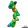
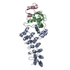
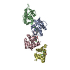
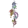
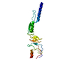
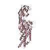
Journal: Proc Natl Acad Sci U S A / Year: 2021 Title: Structural analyses of an RNA stability element interacting with poly(A). Authors: Seyed-Fakhreddin Torabi / Yen-Lin Chen / Kaiming Zhang / Jimin Wang / Suzanne J DeGregorio / Anand T Vaidya / Zhaoming Su / Suzette A Pabit / Wah Chiu / Lois Pollack / Joan A Steitz / Abstract: Cis-acting RNA elements are crucial for the regulation of polyadenylated RNA stability. The element for nuclear expression (ENE) contains a U-rich internal loop flanked by short helices. An ENE ...Cis-acting RNA elements are crucial for the regulation of polyadenylated RNA stability. The element for nuclear expression (ENE) contains a U-rich internal loop flanked by short helices. An ENE stabilizes RNA by sequestering the poly(A) tail via formation of a triplex structure that inhibits a rapid deadenylation-dependent decay pathway. Structure-based bioinformatic studies identified numerous ENE-like elements in evolutionarily diverse genomes, including a subclass containing two ENE motifs separated by a short double-helical region (double ENEs [dENEs]). Here, the structure of a dENE derived from a rice transposable element (TWIFB1) before and after poly(A) binding (∼24 kDa and ∼33 kDa, respectively) is investigated. We combine biochemical structure probing, small angle X-ray scattering (SAXS), and cryo-electron microscopy (cryo-EM) to investigate the dENE structure and its local and global structural changes upon poly(A) binding. Our data reveal 1) the directionality of poly(A) binding to the dENE, and 2) that the dENE-poly(A) interaction involves a motif that protects the 3'-most seven adenylates of the poly(A). Furthermore, we demonstrate that the dENE does not undergo a dramatic global conformational change upon poly(A) binding. These findings are consistent with the recently solved crystal structure of a dENE+poly(A) complex [S.-F. Torabi , 371, eabe6523 (2021)]. Identification of additional modes of poly(A)-RNA interaction opens new venues for better understanding of poly(A) tail biology.
Resolution: 5.6 Å / Resolution method: FSC 0.143 CUT-OFF / Num. of particles: 283486 / Symmetry type: POINT
Atomic model building
Protocol: RIGID BODY FIT
Refinement
Resolution: 5.6→5.6 Å / Cor.coef. Fo:Fc: 0.945 / Cor.coef. Fo:Fc free: 0.885 / SU ML: 110.55 / Cross valid method: THROUGHOUT / ESU R Free: 2.176 Stereochemistry target values: MAXIMUM LIKELIHOOD WITH PHASES Details: HYDROGENS HAVE BEEN ADDED IN THE RIDING POSITIONS
Rfactor
Num. reflection
% reflection
Selection details
Rfree
0.45134
279
6.4 %
RANDOM
Rwork
0.44358
-
-
-
obs
0.4441
4078
100 %
-
Solvent computation
Ion probe radii: 0.8 Å / Shrinkage radii: 0.8 Å / VDW probe radii: 1.2 Å / Solvent model: MASK
 Movie
Movie Controller

 Yorodumi
 Open data
 Basic information
 Components
 Keywords
 Function and homology information
 Authors
 United States, 3items
  Citation
 Structure visualization
 Downloads & links
 Other downloads
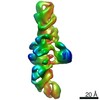


 PDBj
PDBj


 Assembly
 Sample preparation
 Electron microscopy imaging
 FIELD EMISSION GUN / Accelerating voltage: 300 kV / Illumination mode: FLOOD BEAM
 Processing