 Movie
Movie Controller


+ Open data

- Basic information
| Entry | Database: PDB / ID: 7f6v | ||||||
|---|---|---|---|---|---|---|---|
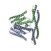
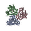
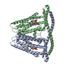
| Title | Cryo-EM structure of the human TACAN channel in a closed state | ||||||
 Components Components | Ion channel TACAN | ||||||
 Keywords Keywords | MEMBRANE PROTEIN / dimer | ||||||
| Function / homology |  Function and homology information Function and homology informationcoenzyme A binding / protein heterooligomerization / nuclear inner membrane / monoatomic ion channel activity / detection of mechanical stimulus involved in sensory perception of pain / fat cell differentiation / antiviral innate immune response / protein homooligomerization / monoatomic ion transmembrane transport / endoplasmic reticulum ...coenzyme A binding / protein heterooligomerization / nuclear inner membrane / monoatomic ion channel activity / detection of mechanical stimulus involved in sensory perception of pain / fat cell differentiation / antiviral innate immune response / protein homooligomerization / monoatomic ion transmembrane transport / endoplasmic reticulum / membrane / plasma membrane Similarity search - Function | ||||||
| Biological species |  Homo sapiens (human) Homo sapiens (human) | ||||||
| Method | ELECTRON MICROSCOPY / single particle reconstruction / cryo EM / Resolution: 3.66 Å | ||||||
 Authors Authors | Chen, X.Z. / Wang, Y.J. / Li, Y. / Yang, X. / Shen, Y.Q. | ||||||
| Funding support |  China, 1items China, 1items
| ||||||
 Citation Citation | Journal: Cell Rep / Year: 2022 Title: Cryo-EM structure of the human TACAN in a closed state. Authors: Xiaozhe Chen / Yaojie Wang / Yang Li / Xuhang Lu / Jianan Chen / Ming Li / Tianlei Wen / Ning Liu / Shenghai Chang / Xing Zhang / Xue Yang / Yuequan Shen / Abstract: TACAN is an ion channel-like protein that may be involved in sensing mechanical pain. Here, we present the cryo-electron microscopic structure of human TACAN (hTACAN). hTACAN forms a dimer in which ...TACAN is an ion channel-like protein that may be involved in sensing mechanical pain. Here, we present the cryo-electron microscopic structure of human TACAN (hTACAN). hTACAN forms a dimer in which each protomer consists of a transmembrane globular domain (TMD) containing six helices and an intracellular domain (ICD) containing two helices. Molecular dynamic simulations suggest that each protomer contains a putative ion conduction pore. A single-point mutation of the key residue Met207 greatly increases membrane pressure-activated currents. In addition, each hTACAN subunit binds one cholesterol molecule. Our data show the molecular assembly of hTACAN and suggest that wild-type hTACAN is in a closed state. | ||||||
| History |
|
- Structure visualization
| Movie |
Movie viewer |
|---|---|
| Structure viewer | Molecule: MolmilJmol/JSmol |
- Downloads & links
-Download
| PDBx/mmCIF format | 7f6v.cif.gz | 123.9 KB | Display | PDBx/mmCIF format |
|---|---|---|---|---|
| PDB format | pdb7f6v.ent.gz | 97.1 KB | Display | PDB format |
| PDBx/mmJSON format | 7f6v.json.gz | Tree view | PDBx/mmJSON format | |
| Others |  Other downloads Other downloads |
-Validation report
| Arichive directory | https://data.pdbj.org/pub/pdb/validation_reports/f6/7f6vftp://data.pdbj.org/pub/pdb/validation_reports/f6/7f6v | HTTPS FTP |
|---|
-Related structure data
| Related structure data | 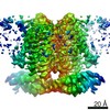 31482MC M: map data used to model this data C: citing same article ( |
|---|---|
| Similar structure data |


-Links
 PDBj
PDBj


- Assembly
| Deposited unit | 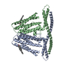
|
|---|---|
| 1 |
|
-Components
| #1: Protein | Mass: 40657.156 Da / Num. of mol.: 2 Source method: isolated from a genetically manipulated source Source: (gene. exp.) Homo sapiens (human) / Gene: TMEM120A, TACAN, TMPIT / Production host: Homo sapiens (human) / References: UniProt: Q9BXJ8#2: Chemical |   Mass: 386.654 Da / Num. of mol.: 2 / Source method: obtained synthetically / Formula: C27H46O Mass: 386.654 Da / Num. of mol.: 2 / Source method: obtained synthetically / Formula: C27H46OHas ligand of interest | N | |
|---|
-Experimental details
-Experiment
| Experiment | Method: ELECTRON MICROSCOPY |
|---|---|
| EM experiment | Aggregation state: PARTICLE / 3D reconstruction method: single particle reconstruction |
- Sample preparation
| Component | Name: Cryo-EM structure of the human TACAN channel in a closed state Type: ORGANELLE OR CELLULAR COMPONENT / Entity ID: #1 / Source: RECOMBINANT |
|---|---|
| Source (natural) | Organism: Homo sapiens (human) |
| Source (recombinant) | Organism: Homo sapiens (human) |
| Buffer solution | pH: 7.5 |
| Specimen | Embedding applied: NO / Shadowing applied: NO / Staining applied: NO / Vitrification applied: YES |
| Specimen support | Grid material: GOLD / Grid type: C-flat-2/1 |
| Vitrification | Instrument: FEI VITROBOT MARK IV / Cryogen name: ETHANE / Humidity: 100 % |
- Electron microscopy imaging
| Experimental equipment |  Model: Titan Krios / Image courtesy: FEI Company |
|---|---|
| Microscopy | Model: FEI TITAN KRIOS |
| Electron gun | Electron source:  FIELD EMISSION GUN / Accelerating voltage: 300 kV / Illumination mode: FLOOD BEAM FIELD EMISSION GUN / Accelerating voltage: 300 kV / Illumination mode: FLOOD BEAM |
| Electron lens | Mode: BRIGHT FIELD / Nominal defocus max: 2000 nm / Nominal defocus min: 1000 nm |
| Specimen holder | Cryogen: NITROGEN / Specimen holder model: FEI TITAN KRIOS AUTOGRID HOLDER |
| Image recording | Electron dose: 56 e/Å2 / Detector mode: SUPER-RESOLUTION / Film or detector model: GATAN K2 SUMMIT (4k x 4k) |
- Processing
| CTF correction | Type: NONE |
|---|---|
| 3D reconstruction | Resolution: 3.66 Å / Resolution method: FSC 0.143 CUT-OFF / Num. of particles: 58843 / Symmetry type: POINT |
| Atomic model building | Protocol: AB INITIO MODEL / Space: REAL |
