 Movie
Movie Controller


+ Open data

- Basic information
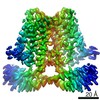
| Entry | Database: PDB / ID: 7f73 | |||||||||||||||||||||||||||||||||||||||||||||
|---|---|---|---|---|---|---|---|---|---|---|---|---|---|---|---|---|---|---|---|---|---|---|---|---|---|---|---|---|---|---|---|---|---|---|---|---|---|---|---|---|---|---|---|---|---|---|
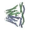
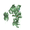
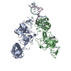
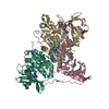
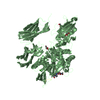
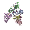
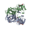
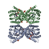
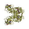
| Title | Cryo-EM structure of human TMEM120B | |||||||||||||||||||||||||||||||||||||||||||||
 Components Components | MCherry fluorescent protein,Transmembrane protein 120B | |||||||||||||||||||||||||||||||||||||||||||||
 Keywords Keywords | MEMBRANE PROTEIN / Membran protein | |||||||||||||||||||||||||||||||||||||||||||||
| Function / homology |  Function and homology information Function and homology informationprotein heterooligomerization / nuclear inner membrane / fat cell differentiation / bioluminescence / generation of precursor metabolites and energy Similarity search - Function | |||||||||||||||||||||||||||||||||||||||||||||
| Biological species |  Anaplasma marginale (bacteria) Anaplasma marginale (bacteria) Homo sapiens (human) Homo sapiens (human) | |||||||||||||||||||||||||||||||||||||||||||||
| Method | ELECTRON MICROSCOPY / single particle reconstruction / cryo EM / Resolution: 4 Å | |||||||||||||||||||||||||||||||||||||||||||||
 Authors Authors | Ke, M. / Wu, J. / Yan, Z. | |||||||||||||||||||||||||||||||||||||||||||||
 Citation Citation | Journal: Cell Discov / Year: 2021 Title: Cryo-EM structures of human TMEM120A and TMEM120B. Authors: Meng Ke / Yue Yu / Changjian Zhao / Shirong Lai / Qiang Su / Weidan Yuan / Lina Yang / Dong Deng / Kun Wu / Weizheng Zeng / Jia Geng / Jianping Wu / Zhen Yan /  | |||||||||||||||||||||||||||||||||||||||||||||
| History |
|
- Structure visualization
| Movie |
Movie viewer |
|---|---|
| Structure viewer | Molecule: MolmilJmol/JSmol |
- Downloads & links
-Download
| PDBx/mmCIF format | 7f73.cif.gz | 128.4 KB | Display | PDBx/mmCIF format |
|---|---|---|---|---|
| PDB format | pdb7f73.ent.gz | 93.2 KB | Display | PDB format |
| PDBx/mmJSON format | 7f73.json.gz | Tree view | PDBx/mmJSON format | |
| Others |  Other downloads Other downloads |
-Validation report
| Arichive directory | https://data.pdbj.org/pub/pdb/validation_reports/f7/7f73ftp://data.pdbj.org/pub/pdb/validation_reports/f7/7f73 | HTTPS FTP |
|---|
-Related structure data
| Related structure data | 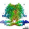 31484MC 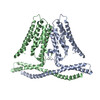 7cxrC C: citing same article ( M: map data used to model this data |
|---|---|
| Similar structure data |
-Links
 PDBj
PDBj

- Assembly
| Deposited unit | 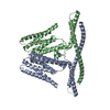
|
|---|---|
| 1 |
|
-Components
| #1: Protein | Mass: 72284.109 Da / Num. of mol.: 2 Source method: isolated from a genetically manipulated source Source: (gene. exp.) Anaplasma marginale (bacteria), (gene. exp.) Homo sapiens (human)Gene: mCherry, TMEM120B / Production host: Homo sapiens (human) / References: UniProt: X5DSL3, UniProt: A0PK00Has protein modification | N | |
|---|
-Experimental details
-Experiment
| Experiment | Method: ELECTRON MICROSCOPY |
|---|---|
| EM experiment | Aggregation state: PARTICLE / 3D reconstruction method: single particle reconstruction |
- Sample preparation
| Component | Name: Dimeric human TMEM120B / Type: COMPLEX / Entity ID: all / Source: RECOMBINANT | ||||||||||||
|---|---|---|---|---|---|---|---|---|---|---|---|---|---|
| Source (natural) |
| ||||||||||||
| Source (recombinant) | Organism: Homo sapiens (human) | ||||||||||||
| Buffer solution | pH: 8 | ||||||||||||
| Specimen | Embedding applied: NO / Shadowing applied: NO / Staining applied: NO / Vitrification applied: YES | ||||||||||||
| Vitrification | Cryogen name: ETHANE |
- Electron microscopy imaging
| Experimental equipment |  Model: Titan Krios / Image courtesy: FEI Company |
|---|---|
| Microscopy | Model: FEI TITAN KRIOS |
| Electron gun | Electron source:  FIELD EMISSION GUN / Accelerating voltage: 300 kV / Illumination mode: FLOOD BEAM FIELD EMISSION GUN / Accelerating voltage: 300 kV / Illumination mode: FLOOD BEAM |
| Electron lens | Mode: BRIGHT FIELD |
| Image recording | Electron dose: 50 e/Å2 / Film or detector model: GATAN K3 (6k x 4k) |
- Processing
| Software | Name: PHENIX / Version: 1.18.1_3865: / Classification: refinement | ||||||||||||||||||||||||
|---|---|---|---|---|---|---|---|---|---|---|---|---|---|---|---|---|---|---|---|---|---|---|---|---|---|
| EM software | Name: PHENIX / Category: model refinement | ||||||||||||||||||||||||
| CTF correction | Type: NONE | ||||||||||||||||||||||||
| 3D reconstruction | Resolution: 4 Å / Resolution method: FSC 0.143 CUT-OFF / Num. of particles: 137998 / Symmetry type: POINT | ||||||||||||||||||||||||
| Refine LS restraints |
|
