 ムービー
ムービー コントローラー


+ データを開く

- 基本情報
基本情報
| 登録情報 | データベース: PDB / ID: 7ell | |||||||||||||||||||||||||||
|---|---|---|---|---|---|---|---|---|---|---|---|---|---|---|---|---|---|---|---|---|---|---|---|---|---|---|---|---|
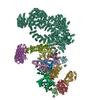
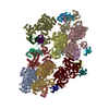
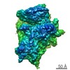
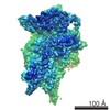
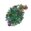
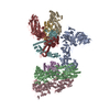
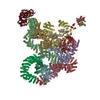
| タイトル | In situ structure of capping enzyme lambda2, penetration protein mu1 of mammalian reovirus capsid asymmetric unit. | |||||||||||||||||||||||||||
 要素 要素 |
| |||||||||||||||||||||||||||
 キーワード キーワード | VIRAL PROTEIN/TRANSFERASE / mammalian reovirus 3 / capping enzyme lambda2 / penetration protein mu1 / VIRUS / VIRAL PROTEIN-TRANSFERASE complex | |||||||||||||||||||||||||||
| 機能・相同性 |  機能・相同性情報 機能・相同性情報host cell surface binding / viral outer capsid / permeabilization of host organelle membrane involved in viral entry into host cell / symbiont entry into host cell via permeabilization of inner membrane / : / mRNA guanylyltransferase activity / mRNA guanylyltransferase / mRNA (guanine-N7)-methyltransferase / mRNA 5'-cap (guanine-N7-)-methyltransferase activity / GTP binding / ATP binding 類似検索 - 分子機能 | |||||||||||||||||||||||||||
| 生物種 |  Mammalian orthoreovirus 3 (ウイルス) Mammalian orthoreovirus 3 (ウイルス) | |||||||||||||||||||||||||||
| 手法 | 電子顕微鏡法 / 単粒子再構成法 / クライオ電子顕微鏡法 / 解像度: 3.8 Å | |||||||||||||||||||||||||||
 データ登録者 データ登録者 | Zhou, Z.H. / Pan, M. | |||||||||||||||||||||||||||
| 資金援助 |  米国, 8件 米国, 8件
| |||||||||||||||||||||||||||
 引用 引用 | ジャーナル: Nat Commun / 年: 2021 タイトル: Asymmetric reconstruction of mammalian reovirus reveals interactions among RNA, transcriptional factor µ2 and capsid proteins. 著者: Muchen Pan / Ana L Alvarez-Cabrera / Joon S Kang / Lihua Wang / Chunhai Fan / Z Hong Zhou /  要旨: Mammalian reovirus (MRV) is the prototypical member of genus Orthoreovirus of family Reoviridae. However, lacking high-resolution structures of its RNA polymerase cofactor μ2 and infectious ...Mammalian reovirus (MRV) is the prototypical member of genus Orthoreovirus of family Reoviridae. However, lacking high-resolution structures of its RNA polymerase cofactor μ2 and infectious particle, limits understanding of molecular interactions among proteins and RNA, and their contributions to virion assembly and RNA transcription. Here, we report the 3.3 Å-resolution asymmetric reconstruction of transcribing MRV and in situ atomic models of its capsid proteins, the asymmetrically attached RNA-dependent RNA polymerase (RdRp) λ3, and RdRp-bound nucleoside triphosphatase μ2 with a unique RNA-binding domain. We reveal molecular interactions among virion proteins and genomic and messenger RNA. Polymerase complexes in three Spinoreovirinae subfamily members are organized with different pseudo-D symmetries to engage their highly diversified genomes. The above interactions and those between symmetry-mismatched receptor-binding σ1 trimers and RNA-capping λ2 pentamers balance competing needs of capsid assembly, external protein removal, and allosteric triggering of endogenous RNA transcription, before, during and after infection, respectively. | |||||||||||||||||||||||||||
| 履歴 |
|
- 構造の表示
構造の表示
| ムービー |
ムービービューア |
|---|---|
| 構造ビューア | 分子: MolmilJmol/JSmol |
- ダウンロードとリンク
-ダウンロード
| PDBx/mmCIF形式 | 7ell.cif.gz | 1.3 MB | 表示 | PDBx/mmCIF形式 |
|---|---|---|---|---|
| PDB形式 | pdb7ell.ent.gz | 1.1 MB | 表示 | PDB形式 |
| PDBx/mmJSON形式 | 7ell.json.gz | ツリー表示 | PDBx/mmJSON形式 | |
| その他 |  その他のダウンロード その他のダウンロード |
-検証レポート
| 文書・要旨 | 7ell_validation.pdf.gz | 1.2 MB | 表示 | wwPDB検証レポート |
|---|---|---|---|---|
| 文書・詳細版 | 7ell_full_validation.pdf.gz | 1.2 MB | 表示 | |
| XML形式データ | 7ell_validation.xml.gz | 196.4 KB | 表示 | |
| CIF形式データ | 7ell_validation.cif.gz | 303.4 KB | 表示 | |
| アーカイブディレクトリ | https://data.pdbj.org/pub/pdb/validation_reports/el/7ellftp://data.pdbj.org/pub/pdb/validation_reports/el/7ell | HTTPS FTP |
-関連構造データ


-リンク
 PDBj
PDBj


- 集合体
集合体
| 登録構造単位 | 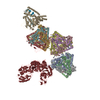
|
|---|---|
| 1 |
|
-要素
| #1: タンパク質・ペプチド | 分子量: 4050.441 Da / 分子数: 10 / 由来タイプ: 天然 / 由来: (天然) Mammalian orthoreovirus 3 (ウイルス) / 参照: UniProt: F1ARM5#2: タンパク質 | 分子量: 72228.719 Da / 分子数: 10 / 由来タイプ: 天然 / 由来: (天然) Mammalian orthoreovirus 3 (ウイルス) / 参照: UniProt: F1ARM5#3: タンパク質 | | 分子量: 143998.625 Da / 分子数: 1 / 由来タイプ: 天然 / 由来: (天然) Mammalian orthoreovirus 3 (ウイルス)参照: UniProt: A0A0B5CUT9, mRNA (guanine-N7)-methyltransferase, mRNA guanylyltransferase #4: 化合物 | ChemComp-MYR /   分子量: 228.371 Da / 分子数: 10 / 由来タイプ: 合成 / 式: C14H28O2 / タイプ: SUBJECT OF INVESTIGATION 分子量: 228.371 Da / 分子数: 10 / 由来タイプ: 合成 / 式: C14H28O2 / タイプ: SUBJECT OF INVESTIGATION研究の焦点であるリガンドがあるか | Y | |
|---|
-実験情報
-実験
| 実験 | 手法: 電子顕微鏡法 |
|---|---|
| EM実験 | 試料の集合状態: PARTICLE / 3次元再構成法: 単粒子再構成法 |
- 試料調製
試料調製
| 構成要素 | 名称: Mammalian orthoreovirus 3 Dearing / タイプ: VIRUS / Entity ID: #3 / 由来: NATURAL |
|---|---|
| 由来(天然) | 生物種: Mammalian orthoreovirus 3 Dearing (ウイルス) |
| ウイルスについての詳細 | 中空か: NO / エンベロープを持つか: NO / 単離: SEROTYPE / タイプ: VIRION |
| 緩衝液 | pH: 7.4 |
| 試料 | 包埋: NO / シャドウイング: NO / 染色: NO / 凍結: YES |
| 急速凍結 | 凍結剤: ETHANE |
- 電子顕微鏡撮影
| 実験機器 |  モデル: Titan Krios / 画像提供: FEI Company |
|---|---|
| 顕微鏡 | モデル: FEI TITAN KRIOS |
| 電子銃 | 電子線源:  FIELD EMISSION GUN / 加速電圧: 300 kV / 照射モード: FLOOD BEAM FIELD EMISSION GUN / 加速電圧: 300 kV / 照射モード: FLOOD BEAM |
| 電子レンズ | モード: BRIGHT FIELD |
| 撮影 | 電子線照射量: 56 e/Å2 フィルム・検出器のモデル: GATAN K2 SUMMIT (4k x 4k) |
- 解析
解析
| CTF補正 | タイプ: PHASE FLIPPING AND AMPLITUDE CORRECTION |
|---|---|
| 3次元再構成 | 解像度: 3.8 Å / 解像度の算出法: FSC 0.143 CUT-OFF / 粒子像の数: 61861 / 対称性のタイプ: POINT |
| 精密化 | 最高解像度: 3.8 Å |
