 Movie
Movie Controller


+ Open data

- Basic information
| Entry | Database: PDB / ID: 7bwm | ||||||||||||||||||||||||
|---|---|---|---|---|---|---|---|---|---|---|---|---|---|---|---|---|---|---|---|---|---|---|---|---|---|
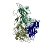
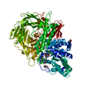
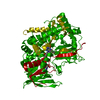
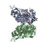
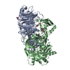
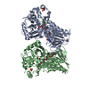
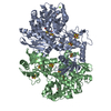
| Title | Cryo-EM structure of the human pathogen Mycoplasma pneumoniae P1 | ||||||||||||||||||||||||
 Components Components | Adhesin P1 | ||||||||||||||||||||||||
 Keywords Keywords | STRUCTURAL PROTEIN / Sialic acid receptor | ||||||||||||||||||||||||
| Function / homology |  Function and homology information Function and homology informationattachment organelle / adhesion of symbiont to microvasculature / cell surface / membrane / plasma membrane Similarity search - Function | ||||||||||||||||||||||||
| Biological species |  Mycoplasma pneumoniae M129 (bacteria) Mycoplasma pneumoniae M129 (bacteria) | ||||||||||||||||||||||||
| Method | ELECTRON MICROSCOPY / single particle reconstruction / cryo EM / Resolution: 2.9 Å | ||||||||||||||||||||||||
 Authors Authors | Kawamoto, A. / Kenri, T. / Namba, K. / Miyata, M. | ||||||||||||||||||||||||
| Funding support |  Japan, 7items Japan, 7items
| ||||||||||||||||||||||||
 Citation Citation | Journal: Nat Commun / Year: 2020 Title: Immunodominant proteins P1 and P40/P90 from human pathogen Mycoplasma pneumoniae. Authors: David Vizarraga / Akihiro Kawamoto / U Matsumoto / Ramiro Illanes / Rosa Pérez-Luque / Jesús Martín / Rocco Mazzolini / Paula Bierge / Oscar Q Pich / Mateu Espasa / Isabel Sanfeliu / ...Authors: David Vizarraga / Akihiro Kawamoto / U Matsumoto / Ramiro Illanes / Rosa Pérez-Luque / Jesús Martín / Rocco Mazzolini / Paula Bierge / Oscar Q Pich / Mateu Espasa / Isabel Sanfeliu / Juliana Esperalba / Miguel Fernández-Huerta / Margot P Scheffer / Jaume Pinyol / Achilleas S Frangakis / Maria Lluch-Senar / Shigetarou Mori / Keigo Shibayama / Tsuyoshi Kenri / Takayuki Kato / Keiichi Namba / Ignacio Fita / Makoto Miyata / David Aparicio /   Abstract: Mycoplasma pneumoniae is a bacterial human pathogen that causes primary atypical pneumonia. M. pneumoniae motility and infectivity are mediated by the immunodominant proteins P1 and P40/P90, which ...Mycoplasma pneumoniae is a bacterial human pathogen that causes primary atypical pneumonia. M. pneumoniae motility and infectivity are mediated by the immunodominant proteins P1 and P40/P90, which form a transmembrane adhesion complex. Here we report the structure of P1, determined by X-ray crystallography and cryo-electron microscopy, and the X-ray structure of P40/P90. Contrary to what had been suggested, the binding site for sialic acid was found in P40/P90 and not in P1. Genetic and clinical variability concentrates on the N-terminal domain surfaces of P1 and P40/P90. Polyclonal antibodies generated against the mostly conserved C-terminal domain of P1 inhibited adhesion of M. pneumoniae, and serology assays with sera from infected patients were positive when tested against this C-terminal domain. P40/P90 also showed strong reactivity against human infected sera. The architectural elements determined for P1 and P40/P90 open new possibilities in vaccine development against M. pneumoniae infections. | ||||||||||||||||||||||||
| History |
|
- Structure visualization
| Movie |
Movie viewer |
|---|---|
| Structure viewer | Molecule: MolmilJmol/JSmol |
- Downloads & links
-Download
| PDBx/mmCIF format | 7bwm.cif.gz | 304.5 KB | Display | PDBx/mmCIF format |
|---|---|---|---|---|
| PDB format | pdb7bwm.ent.gz | 227.9 KB | Display | PDB format |
| PDBx/mmJSON format | 7bwm.json.gz | Tree view | PDBx/mmJSON format | |
| Others |  Other downloads Other downloads |
-Validation report
| Arichive directory | https://data.pdbj.org/pub/pdb/validation_reports/bw/7bwmftp://data.pdbj.org/pub/pdb/validation_reports/bw/7bwm | HTTPS FTP |
|---|
-Related structure data
| Related structure data | 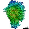 30233MC 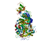 6rc9C  6rj1C  6tlzC 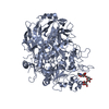 6tm0C M: map data used to model this data C: citing same article ( |
|---|---|
| Similar structure data | |
| EM raw data | EMPIAR-10518 (Title: Cryo-EM structure of immunodominant protein P1 from human pathogen Mycoplasma pneumoniae on graphene oxide grid Data size: 8.0 TB Data #1: Unaligned 136-frame movies of human pathogen Mycoplasma pneumoniae P1 [micrographs - multiframe] Data #2: Unaligned 78-frame movies of human pathogen Mycoplasma pneumoniae P1 [micrographs - multiframe]) |


-Links
 PDBj
PDBj- Assembly
| Deposited unit | 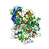
|
|---|---|
| 1 |
|
-Components
| #1: Protein | Mass: 143778.484 Da / Num. of mol.: 1 Source method: isolated from a genetically manipulated source Source: (gene. exp.) Mycoplasma pneumoniae M129 (bacteria) / Strain: M129 / Gene: mgpA, MPN_141, MP013Production host: References: UniProt: P11311 |
|---|
-Experimental details
-Experiment
| Experiment | Method: ELECTRON MICROSCOPY |
|---|---|
| EM experiment | Aggregation state: PARTICLE / 3D reconstruction method: single particle reconstruction |
- Sample preparation
| Component | Name: Nap protein P1 / Type: ORGANELLE OR CELLULAR COMPONENT / Entity ID: all / Source: RECOMBINANT |
|---|---|
| Molecular weight | Value: 170 kDa/nm / Experimental value: NO |
| Source (natural) | Organism: Mycoplasma pneumoniae M129 (bacteria) |
| Source (recombinant) | Organism: |
| Buffer solution | pH: 8 |
| Specimen | Embedding applied: NO / Shadowing applied: NO / Staining applied: NO / Vitrification applied: YES |
| Specimen support | Grid material: COPPER / Grid mesh size: 200 divisions/in. / Grid type: Quantifoil R1.2/1.3 |
| Vitrification | Instrument: FEI VITROBOT MARK IV / Cryogen name: ETHANE / Humidity: 100 % / Chamber temperature: 277 K |
- Electron microscopy imaging
| Experimental equipment |  Model: Titan Krios / Image courtesy: FEI Company |
|---|---|
| Microscopy | Model: FEI TITAN KRIOS |
| Electron gun | Electron source:  FIELD EMISSION GUN / Accelerating voltage: 300 kV / Illumination mode: FLOOD BEAM FIELD EMISSION GUN / Accelerating voltage: 300 kV / Illumination mode: FLOOD BEAM |
| Electron lens | Mode: BRIGHT FIELD / Nominal magnification: 96000 X / Nominal defocus max: 2750 nm / Nominal defocus min: 750 nm / C2 aperture diameter: 50 µm / Alignment procedure: ZEMLIN TABLEAU |
| Specimen holder | Cryogen: NITROGEN / Specimen holder model: FEI TITAN KRIOS AUTOGRID HOLDER / Temperature (max): 78.9 K / Temperature (min): 78.9 K |
| Image recording | Average exposure time: 36.33 sec. / Electron dose: 60 e/Å2 / Detector mode: COUNTING / Film or detector model: FEI FALCON III (4k x 4k) / Num. of real images: 5279 |
| EM imaging optics | Spherical aberration corrector: Microscope was modified with a Cs corrector |
| Image scans | Width: 4096 / Height: 4096 |
- Processing
| Software | Name: PHENIX / Version: 1.17.1_3660: / Classification: refinement | ||||||||||||||||||||||||||||||||||||||||
|---|---|---|---|---|---|---|---|---|---|---|---|---|---|---|---|---|---|---|---|---|---|---|---|---|---|---|---|---|---|---|---|---|---|---|---|---|---|---|---|---|---|
| EM software |
| ||||||||||||||||||||||||||||||||||||||||
| CTF correction | Type: PHASE FLIPPING AND AMPLITUDE CORRECTION | ||||||||||||||||||||||||||||||||||||||||
| Symmetry | Point symmetry: C1 (asymmetric) | ||||||||||||||||||||||||||||||||||||||||
| 3D reconstruction | Resolution: 2.9 Å / Resolution method: FSC 0.143 CUT-OFF / Num. of particles: 68014 / Algorithm: BACK PROJECTION / Symmetry type: POINT | ||||||||||||||||||||||||||||||||||||||||
| Atomic model building | Protocol: RIGID BODY FIT / Space: REAL | ||||||||||||||||||||||||||||||||||||||||
| Atomic model building | PDB-ID: 6RC9 Accession code: 6RC9 / Source name: PDB / Type: experimental model | ||||||||||||||||||||||||||||||||||||||||
| Refine LS restraints |
|
