 Movie
Movie Controller

[English] 日本語
 Yorodumi
- EMDB-30233: Cryo-EM structure of the human pathogen Mycoplasma pneumoniae P1 -
+ Open data

- Basic information
| Entry | Database: EMDB / ID: EMD-30233 | ||||||||||||||||||||||||
|---|---|---|---|---|---|---|---|---|---|---|---|---|---|---|---|---|---|---|---|---|---|---|---|---|---|
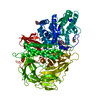
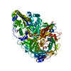
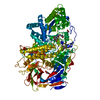
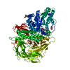
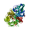
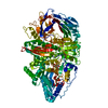
| Title | Cryo-EM structure of the human pathogen Mycoplasma pneumoniae P1 | ||||||||||||||||||||||||
 Map data Map data | |||||||||||||||||||||||||
 Sample Sample |
| ||||||||||||||||||||||||
 Keywords Keywords | Sialic acid receptor / STRUCTURAL PROTEIN | ||||||||||||||||||||||||
| Function / homology |  Function and homology information Function and homology informationattachment organelle / adhesion of symbiont to microvasculature / cell surface / membrane / plasma membrane Similarity search - Function | ||||||||||||||||||||||||
| Biological species |  Mycoplasma pneumoniae M129 (bacteria) Mycoplasma pneumoniae M129 (bacteria) | ||||||||||||||||||||||||
| Method | single particle reconstruction / cryo EM / Resolution: 2.9 Å | ||||||||||||||||||||||||
 Authors Authors | Kawamoto A / Kenri T | ||||||||||||||||||||||||
| Funding support |  Japan, 7 items Japan, 7 items
| ||||||||||||||||||||||||
 Citation Citation | Journal: Nat Commun / Year: 2020 Title: Immunodominant proteins P1 and P40/P90 from human pathogen Mycoplasma pneumoniae. Authors: David Vizarraga / Akihiro Kawamoto / U Matsumoto / Ramiro Illanes / Rosa Pérez-Luque / Jesús Martín / Rocco Mazzolini / Paula Bierge / Oscar Q Pich / Mateu Espasa / Isabel Sanfeliu / ...Authors: David Vizarraga / Akihiro Kawamoto / U Matsumoto / Ramiro Illanes / Rosa Pérez-Luque / Jesús Martín / Rocco Mazzolini / Paula Bierge / Oscar Q Pich / Mateu Espasa / Isabel Sanfeliu / Juliana Esperalba / Miguel Fernández-Huerta / Margot P Scheffer / Jaume Pinyol / Achilleas S Frangakis / Maria Lluch-Senar / Shigetarou Mori / Keigo Shibayama / Tsuyoshi Kenri / Takayuki Kato / Keiichi Namba / Ignacio Fita / Makoto Miyata / David Aparicio /   Abstract: Mycoplasma pneumoniae is a bacterial human pathogen that causes primary atypical pneumonia. M. pneumoniae motility and infectivity are mediated by the immunodominant proteins P1 and P40/P90, which ...Mycoplasma pneumoniae is a bacterial human pathogen that causes primary atypical pneumonia. M. pneumoniae motility and infectivity are mediated by the immunodominant proteins P1 and P40/P90, which form a transmembrane adhesion complex. Here we report the structure of P1, determined by X-ray crystallography and cryo-electron microscopy, and the X-ray structure of P40/P90. Contrary to what had been suggested, the binding site for sialic acid was found in P40/P90 and not in P1. Genetic and clinical variability concentrates on the N-terminal domain surfaces of P1 and P40/P90. Polyclonal antibodies generated against the mostly conserved C-terminal domain of P1 inhibited adhesion of M. pneumoniae, and serology assays with sera from infected patients were positive when tested against this C-terminal domain. P40/P90 also showed strong reactivity against human infected sera. The architectural elements determined for P1 and P40/P90 open new possibilities in vaccine development against M. pneumoniae infections. | ||||||||||||||||||||||||
| History |
|
- Structure visualization
| Movie |
Movie viewer |
|---|---|
| Structure viewer | EM map: SurfViewMolmilJmol/JSmol |
| Supplemental images |

- Downloads & links
-EMDB archive
| Map data | emd_30233.map.gz | 6.5 MB | EMDB map data format | |
|---|---|---|---|---|
| Header (meta data) | emd-30233-v30.xmlemd-30233.xml | 16.8 KB 16.8 KB | Display Display | EMDB header |
| Images | 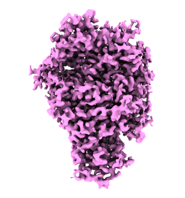 emd_30233.png emd_30233.png | 132.8 KB | ||
| Filedesc metadata | emd-30233.cif.gz | 6.9 KB | ||
| Archive directory |  http://ftp.pdbj.org/pub/emdb/structures/EMD-30233ftp://ftp.pdbj.org/pub/emdb/structures/EMD-30233 http://ftp.pdbj.org/pub/emdb/structures/EMD-30233ftp://ftp.pdbj.org/pub/emdb/structures/EMD-30233 | HTTPS FTP |
-Related structure data
| Related structure data | 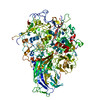 7bwmMC 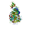 6rc9C  6rj1C  6tlzC 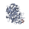 6tm0C M: atomic model generated by this map C: citing same article ( |
|---|---|
| Similar structure data | |
| EM raw data | EMPIAR-10518 (Title: Cryo-EM structure of immunodominant protein P1 from human pathogen Mycoplasma pneumoniae on graphene oxide grid Data size: 8.0 TB Data #1: Unaligned 136-frame movies of human pathogen Mycoplasma pneumoniae P1 [micrographs - multiframe] Data #2: Unaligned 78-frame movies of human pathogen Mycoplasma pneumoniae P1 [micrographs - multiframe]) |


-Links
| EMDB pages | EMDB (EBI/PDBe) / EMDataResource |
|---|
-Map
| File | Download / File: emd_30233.map.gz / Format: CCP4 / Size: 83.7 MB / Type: IMAGE STORED AS FLOATING POINT NUMBER (4 BYTES) | ||||||||||||||||||||||||||||||||||||||||||||||||||||||||||||
|---|---|---|---|---|---|---|---|---|---|---|---|---|---|---|---|---|---|---|---|---|---|---|---|---|---|---|---|---|---|---|---|---|---|---|---|---|---|---|---|---|---|---|---|---|---|---|---|---|---|---|---|---|---|---|---|---|---|---|---|---|---|
| Projections & slices | Image control
Images are generated by Spider. | ||||||||||||||||||||||||||||||||||||||||||||||||||||||||||||
| Voxel size | X=Y=Z: 0.69 Å | ||||||||||||||||||||||||||||||||||||||||||||||||||||||||||||
| Density |
| ||||||||||||||||||||||||||||||||||||||||||||||||||||||||||||
| Symmetry | Space group: 1 | ||||||||||||||||||||||||||||||||||||||||||||||||||||||||||||
| Details | EMDB XML:
CCP4 map header:
| ||||||||||||||||||||||||||||||||||||||||||||||||||||||||||||
 Z (Sec.)
 Y (Row.)
 X (Col.)


-Supplemental data
- Sample components
-Entire : Nap protein P1
| Entire | Name: Nap protein P1 |
|---|---|
| Components |
|
-Supramolecule #1: Nap protein P1
| Supramolecule | Name: Nap protein P1 / type: organelle_or_cellular_component / ID: 1 / Parent: 0 / Macromolecule list: all |
|---|---|
| Source (natural) | Organism: Mycoplasma pneumoniae M129 (bacteria) |
| Molecular weight | Theoretical: 170 kDa/nm |
-Macromolecule #1: Adhesin P1
| Macromolecule | Name: Adhesin P1 / type: protein_or_peptide / ID: 1 / Number of copies: 1 / Enantiomer: LEVO |
|---|---|
| Source (natural) | Organism: Mycoplasma pneumoniae M129 (bacteria) / Strain: M129 |
| Molecular weight | Theoretical: 143.778484 KDa |
| Recombinant expression | Organism: |
| Sequence | String: NAINPRLTPW TYRNTSFSSL PLTGENPGAW ALVRDNSAKG ITAGSGSQQT TYDPTRTEAA LTASTTFALR RYDLAGRALY DLDFSKLNP QTPTRDQTGQ ITFNPFGGFG LSGAAPQQWN EVKNKVPVEV AQDPSNPYRF AVLLVPRSVV YYEQLQRGLG L PQQRTESG ...String: NAINPRLTPW TYRNTSFSSL PLTGENPGAW ALVRDNSAKG ITAGSGSQQT TYDPTRTEAA LTASTTFALR RYDLAGRALY DLDFSKLNP QTPTRDQTGQ ITFNPFGGFG LSGAAPQQWN EVKNKVPVEV AQDPSNPYRF AVLLVPRSVV YYEQLQRGLG L PQQRTESG QNTSTTGAMF GLKVKNAEAD TAKSNEKLQG AEATGSSTTS GSGQSTQRGG SSGDTKVKAL KIEVKKKSDS ED NGQLQLE KNDLANAPIK RSEESGQSVQ LKADDFGTAL SSSGSGGNSN PGSPTPWRPW LATEQIHKDL PKWSASILIL YDA PYARNR TAIDRVDHLD PKAMTANYPP SWRTPKWNHH GLWDWKARDV LLQTTGFFNP RRHPEWFDGG QTVADNEKTG FDVD NSENT KQGFQKEADS DKSAPIALPF EAYFANIGNL TWFGQALLVF GGNGHVTKSA HTAPLSIGVF RVRYNATGTS ATVTG WPYA LLFSGMVNKQ TDGLKDLPFN NNRWFEYVPR MAVAGAKFVG RELVLAGTIT MGDTATVPRL LYDELESNLN LVAQGQ GLL REDLQLFTPY GWANRPDLPI GAWSSSSSSS HNAPYYFHNN PDWQDRPIQN VVDAFIKPWE DKNGKDDAKY IYPYRYS GM WAWQVYNWSN KLTDQPLSAD FVNENAYQPN SLFAAILNPE LLAALPDKVK YGKENEFAAN EYERFNQKLT VAPTQGTN W SHFSPTLSRF STGFNLVGSV LDQVLDYVPW IGNGYRYGNN HRGVDDITAP QTSAGSSSGI STNTSGSRSF LPTFSNIGV GLKANVQATL GGSQTMITGG SPRRTLDQAN LQLWTGAGWR NDKASSGQSD ENHTKFTSAT GMDQQGQSGT SAGNPDSLKQ DNISKSGDS LTTQDGNAID QQEATNYTNL PPNLTPTADW PNALSFTNKN NAQRAQLFLR GLLGSIPVLV NRSGSDSNKF Q ATDQKWSY TDLHSDQTKL NLPAYGEVNG LLNPALVETY FGNTRAGGSG SNTTSSPGIG FKIPEQNNDS KATLITPGLA WT PQDVGNL VVSGTTVSFQ LGGWLVTFTD FVKPRAGYLG LQLTGLDASD ATQRALIWAP RPWAAFRGSW VNRLGRVESV WDL KGVWAD QAQSDSQGST TTATRNALPE HPNALAFQVS VVEASAYKPN TSSGQTQSTN SSPYLHLVKP KKVTQSDKLD DDLK NLLDP NQVRTKLRQS FGTDHSTQPQ PQSLKTTTPV FGTSSGNLSS VLSGGGAGGG SSGSGQSGVD LSPVEKVSGW LVGQL PSTS DGNTSSTNNL APNTNTGNDV VGVGRLSESN AAKMNDDVDG IVRT UniProtKB: Adhesin P1 |
-Experimental details
-Structure determination
| Method | cryo EM |
|---|---|
 Processing Processing | single particle reconstruction |
| Aggregation state | particle |
-Sample preparation
| Buffer | pH: 8 |
|---|---|
| Grid | Model: Quantifoil R1.2/1.3 / Material: COPPER / Mesh: 200 / Support film - Material: GRAPHENE OXIDE / Support film - topology: CONTINUOUS |
| Vitrification | Cryogen name: ETHANE / Chamber humidity: 100 % / Chamber temperature: 277 K / Instrument: FEI VITROBOT MARK IV |
- Electron microscopy
| Microscope | FEI TITAN KRIOS |
|---|---|
| Temperature | Min: 78.9 K / Max: 78.9 K |
| Specialist optics | Spherical aberration corrector: Microscope was modified with a Cs corrector |
| Image recording | Film or detector model: FEI FALCON III (4k x 4k) / Detector mode: COUNTING / Digitization - Dimensions - Width: 4096 pixel / Digitization - Dimensions - Height: 4096 pixel / Number real images: 5279 / Average exposure time: 36.33 sec. / Average electron dose: 60.0 e/Å2 |
| Electron beam | Acceleration voltage: 300 kV / Electron source:  FIELD EMISSION GUN FIELD EMISSION GUN |
| Electron optics | C2 aperture diameter: 50.0 µm / Illumination mode: FLOOD BEAM / Imaging mode: BRIGHT FIELD / Nominal defocus max: 2.75 µm / Nominal defocus min: 0.75 µm / Nominal magnification: 96000 |
| Sample stage | Specimen holder model: FEI TITAN KRIOS AUTOGRID HOLDER / Cooling holder cryogen: NITROGEN |
| Experimental equipment |  Model: Titan Krios / Image courtesy: FEI Company |
