 Movie
Movie Controller


+ Open data

- Basic information
| Entry | Database: PDB / ID: 6y7e | |||||||||
|---|---|---|---|---|---|---|---|---|---|---|
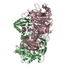
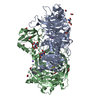
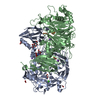
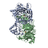
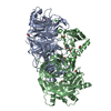
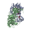
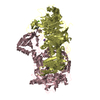
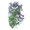
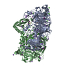
| Title | Pseudomonas stutzeri nitrous oxide reductase mutant, H494A | |||||||||
 Components Components | Nitrous-oxide reductase | |||||||||
 Keywords Keywords | OXIDOREDUCTASE / periplasmic copper-binding protein | |||||||||
| Function / homology |  Function and homology information Function and homology informationnitrous-oxide reductase / nitrous-oxide reductase activity / copper ion import / denitrification pathway / cytochrome-c oxidase activity / periplasmic space / copper ion binding / calcium ion binding / membrane Similarity search - Function | |||||||||
| Biological species |  Pseudomonas stutzeri (bacteria) Pseudomonas stutzeri (bacteria) | |||||||||
| Method |  X-RAY DIFFRACTION / SYNCHROTRON / MOLECULAR REPLACEMENT / Resolution: 1.6 Å X-RAY DIFFRACTION / SYNCHROTRON / MOLECULAR REPLACEMENT / Resolution: 1.6 Å | |||||||||
 Authors Authors | Zhang, L. / Kroneck, P.M.H. / Einsle, O. | |||||||||
| Funding support |  Germany, 2items Germany, 2items
| |||||||||
 Citation Citation | Journal: Chem Sci / Year: 2021 Title: A [3Cu:2S] cluster provides insight into the assembly and function of the Cu Z site of nitrous oxide reductase. Authors: Zhang, L. / Bill, E. / Kroneck, P.M.H. / Einsle, O. | |||||||||
| History |
|
- Structure visualization
| Structure viewer | Molecule: MolmilJmol/JSmol |
|---|
- Downloads & links
-Download
| PDBx/mmCIF format | 6y7e.cif.gz | 493.8 KB | Display | PDBx/mmCIF format |
|---|---|---|---|---|
| PDB format | pdb6y7e.ent.gz | 399.9 KB | Display | PDB format |
| PDBx/mmJSON format | 6y7e.json.gz | Tree view | PDBx/mmJSON format | |
| Others |  Other downloads Other downloads |
-Validation report
| Arichive directory | https://data.pdbj.org/pub/pdb/validation_reports/y7/6y7eftp://data.pdbj.org/pub/pdb/validation_reports/y7/6y7e | HTTPS FTP |
|---|
-Related structure data
| Related structure data | 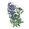 6y6yC 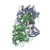 6y71C  6y72C 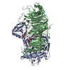 6y77C  6y7dC  7aqaC  3sbqS S: Starting model for refinement C: citing same article ( |
|---|---|
| Similar structure data |

-Links
 PDBj
PDBj


- Assembly
| Deposited unit | 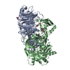
| ||||||||
|---|---|---|---|---|---|---|---|---|---|
| 1 |
| ||||||||
| Unit cell |
|
-Components
-Protein , 1 types, 2 molecules AB
| #1: Protein | Mass: 71891.484 Da / Num. of mol.: 2 Source method: isolated from a genetically manipulated source Source: (gene. exp.) Pseudomonas stutzeri (bacteria) / Gene: nosZ / Production host: |
|---|
-Non-polymers , 9 types, 1112 molecules 


| #2: Chemical | ChemComp-FMT /  Mass: 46.025 Da / Num. of mol.: 4 / Source method: obtained synthetically / Formula: CH2O2 Mass: 46.025 Da / Num. of mol.: 4 / Source method: obtained synthetically / Formula: CH2O2#3: Chemical |  Mass: 65.409 Da / Num. of mol.: 3 / Source method: obtained synthetically / Formula: Zn / Feature type: SUBJECT OF INVESTIGATION Mass: 65.409 Da / Num. of mol.: 3 / Source method: obtained synthetically / Formula: Zn / Feature type: SUBJECT OF INVESTIGATION#4: Chemical | ChemComp-TRS / |  Mass: 122.143 Da / Num. of mol.: 1 / Source method: obtained synthetically / Formula: C4H12NO3 / Comment: pH buffer*YM Mass: 122.143 Da / Num. of mol.: 1 / Source method: obtained synthetically / Formula: C4H12NO3 / Comment: pH buffer*YM#5: Chemical |  Mass: 282.334 Da / Num. of mol.: 2 / Source method: obtained synthetically / Formula: C11H26N2O6 / Comment: pH buffer*YM Mass: 282.334 Da / Num. of mol.: 2 / Source method: obtained synthetically / Formula: C11H26N2O6 / Comment: pH buffer*YM#6: Chemical |  Mass: 22.990 Da / Num. of mol.: 2 / Source method: obtained synthetically / Formula: Na Mass: 22.990 Da / Num. of mol.: 2 / Source method: obtained synthetically / Formula: Na#7: Chemical |  Mass: 35.453 Da / Num. of mol.: 2 / Source method: obtained synthetically / Formula: Cl Mass: 35.453 Da / Num. of mol.: 2 / Source method: obtained synthetically / Formula: Cl#8: Chemical |  Mass: 127.092 Da / Num. of mol.: 2 / Source method: obtained synthetically / Formula: Cu2 / Feature type: SUBJECT OF INVESTIGATION Mass: 127.092 Da / Num. of mol.: 2 / Source method: obtained synthetically / Formula: Cu2 / Feature type: SUBJECT OF INVESTIGATION#9: Chemical | ChemComp-CA / |  Mass: 40.078 Da / Num. of mol.: 1 / Source method: obtained synthetically / Formula: Ca Mass: 40.078 Da / Num. of mol.: 1 / Source method: obtained synthetically / Formula: Ca#10: Water | ChemComp-HOH / | Mass: 18.015 Da / Num. of mol.: 1095 / Source method: isolated from a natural source / Formula: H2O |
|---|
-Details
| Has ligand of interest | Y |
|---|
-Experimental details
-Experiment
| Experiment | Method: X-RAY DIFFRACTION / Number of used crystals: 1 |
|---|
- Sample preparation
| Crystal | Density Matthews: 2 Å3/Da / Density % sol: 38.46 % |
|---|---|
| Crystal grow | Temperature: 293 K / Method: vapor diffusion, sitting drop Details: 0.1 M bis-tris propane buffer at pH 8.5, 0.1 M sodium formate, 0.1 M sodium chloride, and 25% (w/v) of a medium molecular weight (MMW) polyethylene glycol mixture (PEG 2K, 3350, 4K and 5K MME) |
-Data collection
| Diffraction | Mean temperature: 100 K / Serial crystal experiment: N |
|---|---|
| Diffraction source | Source: SYNCHROTRON / Site: SLS  / Beamline: X06DA / Wavelength: 1.36999 Å / Beamline: X06DA / Wavelength: 1.36999 Å |
| Detector | Type: DECTRIS PILATUS 2M-F / Detector: PIXEL / Date: May 5, 2018 |
| Radiation | Protocol: SINGLE WAVELENGTH / Monochromatic (M) / Laue (L): M / Scattering type: x-ray |
| Radiation wavelength | Wavelength: 1.36999 Å / Relative weight: 1 |
| Reflection | Resolution: 1.6→108.3 Å / Num. obs: 110732 / % possible obs: 91.7 % / Redundancy: 7 % / CC1/2: 0.999 / Net I/σ(I): 15.3 |
| Reflection shell | Resolution: 1.6→1.74 Å / Num. unique obs: 5531 / CC1/2: 0.624 |
- Processing
| Software |
| |||||||||||||||||||||||||||||||||||||||||||||||||||||||||||||||||||||||||||
|---|---|---|---|---|---|---|---|---|---|---|---|---|---|---|---|---|---|---|---|---|---|---|---|---|---|---|---|---|---|---|---|---|---|---|---|---|---|---|---|---|---|---|---|---|---|---|---|---|---|---|---|---|---|---|---|---|---|---|---|---|---|---|---|---|---|---|---|---|---|---|---|---|---|---|---|---|
| Refinement | Method to determine structure: MOLECULAR REPLACEMENT Starting model: 3sbq Resolution: 1.6→108.3 Å / Cross valid method: FREE R-VALUE
| |||||||||||||||||||||||||||||||||||||||||||||||||||||||||||||||||||||||||||
| Displacement parameters | Biso max: 168.18 Å2 / Biso min: 10.88 Å2 | |||||||||||||||||||||||||||||||||||||||||||||||||||||||||||||||||||||||||||
| Refinement step | Cycle: final / Resolution: 1.6→108.3 Å
| |||||||||||||||||||||||||||||||||||||||||||||||||||||||||||||||||||||||||||
| Refinement TLS params. | Method: refined / Refine-ID: X-RAY DIFFRACTION
| |||||||||||||||||||||||||||||||||||||||||||||||||||||||||||||||||||||||||||
| Refinement TLS group |
|
