 Movie
Movie Controller

[English] 日本語
 Yorodumi
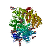
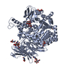
- PDB-7p7p: Crystal structure of ERAP2 aminopeptidase in complex with phosphi... -
+ Open data

- Basic information
| Entry | Database: PDB / ID: 7p7p | ||||||
|---|---|---|---|---|---|---|---|
| Title | Crystal structure of ERAP2 aminopeptidase in complex with phosphinic pseudotripeptide((1R)-1-Amino-3-phenylpropyl){(2S)-3-[((2S)-1-amino-1-oxo-3-phenylpropan-2-yl)amino]-2-{[3-(2-hydroxyphenyl)-isoxazol-5-yl]methyl}-3-oxopropyl}phosphinic acid | ||||||
 Components Components | Endoplasmic reticulum aminopeptidase 2 | ||||||
 Keywords Keywords | HYDROLASE / ERAP2 / transition state analogues / antigen presentation / phosphinic pseudotripeptides / aminopeptidase | ||||||
| Function / homology |  Function and homology information Function and homology informationHydrolases; Acting on peptide bonds (peptidases); Aminopeptidases / antigen processing and presentation of peptide antigen via MHC class I / peptide catabolic process / metalloaminopeptidase activity / aminopeptidase activity / Antigen Presentation: Folding, assembly and peptide loading of class I MHC / peptide binding / antigen processing and presentation of endogenous peptide antigen via MHC class I / regulation of blood pressure / metallopeptidase activity ...Hydrolases; Acting on peptide bonds (peptidases); Aminopeptidases / antigen processing and presentation of peptide antigen via MHC class I / peptide catabolic process / metalloaminopeptidase activity / aminopeptidase activity / Antigen Presentation: Folding, assembly and peptide loading of class I MHC / peptide binding / antigen processing and presentation of endogenous peptide antigen via MHC class I / regulation of blood pressure / metallopeptidase activity / adaptive immune response / endopeptidase activity / endoplasmic reticulum lumen / endoplasmic reticulum membrane / proteolysis / extracellular space / zinc ion binding / membrane / cytoplasm Similarity search - Function | ||||||
| Biological species |  Homo sapiens (human) Homo sapiens (human) | ||||||
| Method |  X-RAY DIFFRACTION / SYNCHROTRON / MOLECULAR REPLACEMENT / Resolution: 3 Å X-RAY DIFFRACTION / SYNCHROTRON / MOLECULAR REPLACEMENT / Resolution: 3 Å | ||||||
 Authors Authors | Giastas, P. / Stratikos, E. / Mpakali, A. | ||||||
| Funding support | European Union, 1items
| ||||||
 Citation Citation | Journal: Acs Med.Chem.Lett. / Year: 2022 Title: Inhibitor-Dependent Usage of the S1' Specificity Pocket of ER Aminopeptidase 2. Authors: Mpakali, A. / Georgiadis, D. / Stratikos, E. / Giastas, P. | ||||||
| History |
|
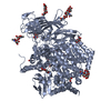
- Structure visualization
| Structure viewer | Molecule: MolmilJmol/JSmol |
|---|
- Downloads & links
-Download
| PDBx/mmCIF format | 7p7p.cif.gz | 913.3 KB | Display | PDBx/mmCIF format |
|---|---|---|---|---|
| PDB format | pdb7p7p.ent.gz | 633.7 KB | Display | PDB format |
| PDBx/mmJSON format | 7p7p.json.gz | Tree view | PDBx/mmJSON format | |
| Others |  Other downloads Other downloads |
-Validation report
| Summary document | 7p7p_validation.pdf.gz | 3.2 MB | Display | wwPDB validaton report |
|---|---|---|---|---|
| Full document | 7p7p_full_validation.pdf.gz | 3.2 MB | Display | |
| Data in XML | 7p7p_validation.xml.gz | 72.5 KB | Display | |
| Data in CIF | 7p7p_validation.cif.gz | 98.1 KB | Display | |
| Arichive directory | https://data.pdbj.org/pub/pdb/validation_reports/p7/7p7pftp://data.pdbj.org/pub/pdb/validation_reports/p7/7p7p | HTTPS FTP |
-Related structure data
| Related structure data | 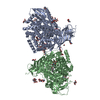 7pfsC  5ab0S S: Starting model for refinement C: citing same article ( |
|---|---|
| Similar structure data |


-Links
 PDBj
PDBj


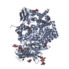
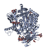
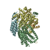
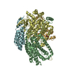
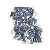
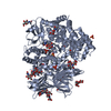
- Assembly
| Deposited unit | 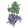
| ||||||||||||
|---|---|---|---|---|---|---|---|---|---|---|---|---|---|
| 1 | 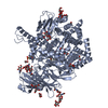
| ||||||||||||
| 2 | 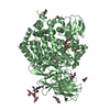
| ||||||||||||
| Unit cell |
|
-Components
-Protein , 1 types, 2 molecules AB
| #1: Protein | Mass: 110872.672 Da / Num. of mol.: 2 / Mutation: K392N Source method: isolated from a genetically manipulated source Source: (gene. exp.) Homo sapiens (human) / Gene: ERAP2, LRAP / Production host:  Trichoplusia ni (cabbage looper) Trichoplusia ni (cabbage looper)References: UniProt: Q6P179, Hydrolases; Acting on peptide bonds (peptidases); Aminopeptidases |
|---|
-Sugars , 6 types, 17 molecules 
| #2: Polysaccharide | alpha-D-mannopyranose-(1-3)-beta-D-mannopyranose-(1-4)-2-acetamido-2-deoxy-beta-D-glucopyranose-(1- ...alpha-D-mannopyranose-(1-3)-beta-D-mannopyranose-(1-4)-2-acetamido-2-deoxy-beta-D-glucopyranose-(1-4)-2-acetamido-2-deoxy-beta-D-glucopyranose Source method: isolated from a genetically manipulated source | ||||||
|---|---|---|---|---|---|---|---|
| #3: Polysaccharide | alpha-D-mannopyranose-(1-6)-beta-D-mannopyranose-(1-4)-2-acetamido-2-deoxy-beta-D-glucopyranose-(1- ...alpha-D-mannopyranose-(1-6)-beta-D-mannopyranose-(1-4)-2-acetamido-2-deoxy-beta-D-glucopyranose-(1-4)-2-acetamido-2-deoxy-beta-D-glucopyranose Source method: isolated from a genetically manipulated source | ||||||
| #4: Polysaccharide | Source method: isolated from a genetically manipulated source #5: Polysaccharide | beta-D-mannopyranose-(1-4)-2-acetamido-2-deoxy-beta-D-glucopyranose-(1-4)-2-acetamido-2-deoxy-beta- ...beta-D-mannopyranose-(1-4)-2-acetamido-2-deoxy-beta-D-glucopyranose-(1-4)-2-acetamido-2-deoxy-beta-D-glucopyranose | Source method: isolated from a genetically manipulated source #6: Polysaccharide | alpha-D-mannopyranose-(1-3)-[alpha-D-mannopyranose-(1-6)]beta-D-mannopyranose-(1-4)-2-acetamido-2- ...alpha-D-mannopyranose-(1-3)-[alpha-D-mannopyranose-(1-6)]beta-D-mannopyranose-(1-4)-2-acetamido-2-deoxy-beta-D-glucopyranose-(1-4)-2-acetamido-2-deoxy-beta-D-glucopyranose | Source method: isolated from a genetically manipulated source #8: Sugar | ChemComp-NAG /  Type: D-saccharide, beta linking / Mass: 221.208 Da / Num. of mol.: 10 Type: D-saccharide, beta linking / Mass: 221.208 Da / Num. of mol.: 10Source method: isolated from a genetically manipulated source Formula: C8H15NO6 |
-Non-polymers , 9 types, 174 molecules 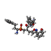


| #7: Chemical |  Mass: 590.607 Da / Num. of mol.: 2 Mass: 590.607 Da / Num. of mol.: 2Source method: isolated from a genetically manipulated source Formula: C31H35N4O6P #9: Chemical |  Mass: 106.120 Da / Num. of mol.: 2 Mass: 106.120 Da / Num. of mol.: 2Source method: isolated from a genetically manipulated source Formula: C4H10O3 #10: Chemical | ChemComp-PGE / |  Mass: 150.173 Da / Num. of mol.: 1 Mass: 150.173 Da / Num. of mol.: 1Source method: isolated from a genetically manipulated source Formula: C6H14O4 #11: Chemical | ChemComp-MES / |  Mass: 195.237 Da / Num. of mol.: 1 / Source method: obtained synthetically / Formula: C6H13NO4S / Comment: pH buffer*YM Mass: 195.237 Da / Num. of mol.: 1 / Source method: obtained synthetically / Formula: C6H13NO4S / Comment: pH buffer*YM#12: Chemical | ChemComp-EDO /  Mass: 62.068 Da / Num. of mol.: 7 / Source method: obtained synthetically / Formula: C2H6O2 Mass: 62.068 Da / Num. of mol.: 7 / Source method: obtained synthetically / Formula: C2H6O2#13: Chemical |  Mass: 76.094 Da / Num. of mol.: 2 / Source method: obtained synthetically / Formula: C3H8O2 Mass: 76.094 Da / Num. of mol.: 2 / Source method: obtained synthetically / Formula: C3H8O2#14: Chemical | ChemComp-IMD / |  Mass: 69.085 Da / Num. of mol.: 1 / Source method: obtained synthetically / Formula: C3H5N2 Mass: 69.085 Da / Num. of mol.: 1 / Source method: obtained synthetically / Formula: C3H5N2#15: Chemical |  Mass: 65.409 Da / Num. of mol.: 2 / Source method: obtained synthetically / Formula: Zn Mass: 65.409 Da / Num. of mol.: 2 / Source method: obtained synthetically / Formula: Zn#16: Water | ChemComp-HOH / | Mass: 18.015 Da / Num. of mol.: 156 / Source method: isolated from a natural source / Formula: H2O |
|---|
-Details
| Has ligand of interest | N |
|---|
-Experimental details
-Experiment
| Experiment | Method: X-RAY DIFFRACTION / Number of used crystals: 1 |
|---|
- Sample preparation
| Crystal | Density Matthews: 3.03 Å3/Da / Density % sol: 58.08 % |
|---|---|
| Crystal grow | Temperature: 277.15 K / Method: vapor diffusion, hanging drop / pH: 6.5 Details: 10% w/v PEG 8000, 20% v/v ethylene glycol, 0.02M of each alcohol (0.2M 1,6-hexanediol, 0.2M 1-butanol, 0.2M (RS)-1,2-propanediol, 0.2M 2-propanol, 0.2M 1,4-butanediol, 0.2M 1,3-propanediol), ...Details: 10% w/v PEG 8000, 20% v/v ethylene glycol, 0.02M of each alcohol (0.2M 1,6-hexanediol, 0.2M 1-butanol, 0.2M (RS)-1,2-propanediol, 0.2M 2-propanol, 0.2M 1,4-butanediol, 0.2M 1,3-propanediol), 0.1M MES/imidazole pH 6.5 |
-Data collection
| Diffraction | Mean temperature: 100 K / Serial crystal experiment: N |
|---|---|
| Diffraction source | Source: SYNCHROTRON / Site: PETRA III, EMBL c/o DESY  / Beamline: P13 (MX1) / Wavelength: 0.9763 Å / Beamline: P13 (MX1) / Wavelength: 0.9763 Å |
| Detector | Type: DECTRIS PILATUS 6M / Detector: PIXEL / Date: Aug 12, 2019 |
| Radiation | Protocol: SINGLE WAVELENGTH / Monochromatic (M) / Laue (L): M / Scattering type: x-ray |
| Radiation wavelength | Wavelength: 0.9763 Å / Relative weight: 1 |
| Reflection | Resolution: 3→50.19 Å / Num. obs: 48793 / % possible obs: 94.5 % / Redundancy: 2 % / Biso Wilson estimate: 79.48 Å2 / CC1/2: 0.997 / Rmerge(I) obs: 0.05698 / Net I/σ(I): 10.98 |
| Reflection shell | Resolution: 3→3.107 Å / Rmerge(I) obs: 0.4295 / Mean I/σ(I) obs: 1.46 / Num. unique obs: 2984 / CC1/2: 0.607 |
- Processing
| Software |
| |||||||||||||||||||||||||||||||||||||||||||||||||||||||||||||||||||||||||||||||||||||||||||||||||||||||||||||||||||||||
|---|---|---|---|---|---|---|---|---|---|---|---|---|---|---|---|---|---|---|---|---|---|---|---|---|---|---|---|---|---|---|---|---|---|---|---|---|---|---|---|---|---|---|---|---|---|---|---|---|---|---|---|---|---|---|---|---|---|---|---|---|---|---|---|---|---|---|---|---|---|---|---|---|---|---|---|---|---|---|---|---|---|---|---|---|---|---|---|---|---|---|---|---|---|---|---|---|---|---|---|---|---|---|---|---|---|---|---|---|---|---|---|---|---|---|---|---|---|---|---|---|
| Refinement | Method to determine structure: MOLECULAR REPLACEMENT Starting model: 5AB0 Resolution: 3→50.19 Å / SU ML: 0.4145 / Cross valid method: FREE R-VALUE / σ(F): 1.38 / Phase error: 25.7533 Stereochemistry target values: GeoStd + Monomer Library + CDL v1.2
| |||||||||||||||||||||||||||||||||||||||||||||||||||||||||||||||||||||||||||||||||||||||||||||||||||||||||||||||||||||||
| Solvent computation | Shrinkage radii: 0.9 Å / VDW probe radii: 1.11 Å / Solvent model: FLAT BULK SOLVENT MODEL / Bsol: 78.9 Å2 / ksol: 0.29 e/Å3 | |||||||||||||||||||||||||||||||||||||||||||||||||||||||||||||||||||||||||||||||||||||||||||||||||||||||||||||||||||||||
| Displacement parameters | Biso mean: 92.48 Å2 | |||||||||||||||||||||||||||||||||||||||||||||||||||||||||||||||||||||||||||||||||||||||||||||||||||||||||||||||||||||||
| Refinement step | Cycle: LAST / Resolution: 3→50.19 Å
| |||||||||||||||||||||||||||||||||||||||||||||||||||||||||||||||||||||||||||||||||||||||||||||||||||||||||||||||||||||||
| Refine LS restraints |
| |||||||||||||||||||||||||||||||||||||||||||||||||||||||||||||||||||||||||||||||||||||||||||||||||||||||||||||||||||||||
| LS refinement shell |
| |||||||||||||||||||||||||||||||||||||||||||||||||||||||||||||||||||||||||||||||||||||||||||||||||||||||||||||||||||||||
| Refinement TLS params. | Method: refined / Origin x: 19.048197273 Å / Origin y: 3.00992509179 Å / Origin z: 30.1524299837 Å
| |||||||||||||||||||||||||||||||||||||||||||||||||||||||||||||||||||||||||||||||||||||||||||||||||||||||||||||||||||||||
| Refinement TLS group | Selection details: all |
