 Movie
Movie Controller

[English] 日本語
 Yorodumi
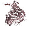
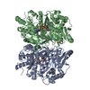
- PDB-5j6s: Crystal structure of Endoplasmic Reticulum Aminopeptidase 2 (ERAP... -
+ Open data

- Basic information
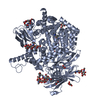
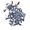
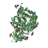
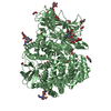
| Entry | Database: PDB / ID: 5j6s | |||||||||
|---|---|---|---|---|---|---|---|---|---|---|
| Title | Crystal structure of Endoplasmic Reticulum Aminopeptidase 2 (ERAP2) in complex with a hydroxamic derivative ligand | |||||||||
 Components Components | Endoplasmic reticulum aminopeptidase 2 | |||||||||
 Keywords Keywords | HYDROLASE / Endoplasmic reticulum / aminopeptidase / Zn binding metallopeptidase | |||||||||
| Function / homology |  Function and homology information Function and homology informationHydrolases; Acting on peptide bonds (peptidases); Aminopeptidases / peptide catabolic process / metalloaminopeptidase activity / aminopeptidase activity / antigen processing and presentation of peptide antigen via MHC class I / Antigen Presentation: Folding, assembly and peptide loading of class I MHC / antigen processing and presentation of endogenous peptide antigen via MHC class I / regulation of blood pressure / metallopeptidase activity / endopeptidase activity ...Hydrolases; Acting on peptide bonds (peptidases); Aminopeptidases / peptide catabolic process / metalloaminopeptidase activity / aminopeptidase activity / antigen processing and presentation of peptide antigen via MHC class I / Antigen Presentation: Folding, assembly and peptide loading of class I MHC / antigen processing and presentation of endogenous peptide antigen via MHC class I / regulation of blood pressure / metallopeptidase activity / endopeptidase activity / adaptive immune response / endoplasmic reticulum lumen / endoplasmic reticulum membrane / proteolysis / zinc ion binding Similarity search - Function | |||||||||
| Biological species |  Homo sapiens (human) Homo sapiens (human) | |||||||||
| Method |  X-RAY DIFFRACTION / SYNCHROTRON / MOLECULAR REPLACEMENT / Resolution: 2.8 Å X-RAY DIFFRACTION / SYNCHROTRON / MOLECULAR REPLACEMENT / Resolution: 2.8 Å | |||||||||
 Authors Authors | Saridakis, E. / Giastas, P. / Mpakali, A. / Deprez-Poulain, R. / Stratikos, E. | |||||||||
| Funding support |  Greece, 1items Greece, 1items
| |||||||||
 Citation Citation | Journal: ACS Med Chem Lett / Year: 2017 Title: Crystal Structures of ERAP2 Complexed with Inhibitors Reveal Pharmacophore Requirements for Optimizing Inhibitor Potency. Authors: Mpakali, A. / Giastas, P. / Deprez-Poulain, R. / Papakyriakou, A. / Koumantou, D. / Gealageas, R. / Tsoukalidou, S. / Vourloumis, D. / Mavridis, I.M. / Stratikos, E. / Saridakis, E. | |||||||||
| History |
|
- Structure visualization
| Structure viewer | Molecule: MolmilJmol/JSmol |
|---|
- Downloads & links
-Download
| PDBx/mmCIF format | 5j6s.cif.gz | 386.3 KB | Display | PDBx/mmCIF format |
|---|---|---|---|---|
| PDB format | pdb5j6s.ent.gz | 311.3 KB | Display | PDB format |
| PDBx/mmJSON format | 5j6s.json.gz | Tree view | PDBx/mmJSON format | |
| Others |  Other downloads Other downloads |
-Validation report
| Arichive directory | https://data.pdbj.org/pub/pdb/validation_reports/j6/5j6sftp://data.pdbj.org/pub/pdb/validation_reports/j6/5j6s | HTTPS FTP |
|---|
-Related structure data
| Related structure data |  5k1vC  5ab0S S: Starting model for refinement C: citing same article ( |
|---|---|
| Similar structure data |


-Links
 PDBj
PDBj- Assembly
| Deposited unit | 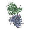
| ||||||||
|---|---|---|---|---|---|---|---|---|---|
| 1 | 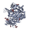
| ||||||||
| 2 | 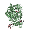
| ||||||||
| Unit cell |
|
-Components
-Protein , 1 types, 2 molecules AB
| #1: Protein | Mass: 111563.422 Da / Num. of mol.: 2 Source method: isolated from a genetically manipulated source Source: (gene. exp.) Homo sapiens (human) / Gene: ERAP2, LRAP / Production host:  Trichoplusia ni (cabbage looper) Trichoplusia ni (cabbage looper)References: UniProt: Q6P179, Hydrolases; Acting on peptide bonds (peptidases); Aminopeptidases |
|---|
-Sugars , 4 types, 16 molecules 
| #2: Polysaccharide | | #3: Polysaccharide | #4: Polysaccharide | alpha-D-mannopyranose-(1-3)-[alpha-D-mannopyranose-(1-6)]beta-D-mannopyranose-(1-4)-2-acetamido-2- ...alpha-D-mannopyranose-(1-3)-[alpha-D-mannopyranose-(1-6)]beta-D-mannopyranose-(1-4)-2-acetamido-2-deoxy-beta-D-glucopyranose-(1-4)-2-acetamido-2-deoxy-beta-D-glucopyranose | Source method: isolated from a genetically manipulated source #6: Sugar | ChemComp-NAG /  Type: D-saccharide, beta linking / Mass: 221.208 Da / Num. of mol.: 11 / Source method: obtained synthetically / Formula: C8H15NO6 Type: D-saccharide, beta linking / Mass: 221.208 Da / Num. of mol.: 11 / Source method: obtained synthetically / Formula: C8H15NO6 |
|---|
-Non-polymers , 3 types, 53 molecules 
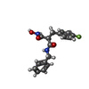

| #5: Chemical |  Mass: 65.409 Da / Num. of mol.: 2 / Source method: obtained synthetically / Formula: Zn Mass: 65.409 Da / Num. of mol.: 2 / Source method: obtained synthetically / Formula: Zn#7: Chemical |  Mass: 316.327 Da / Num. of mol.: 2 Mass: 316.327 Da / Num. of mol.: 2Source method: isolated from a genetically manipulated source Formula: C17H17FN2O3 #8: Water | ChemComp-HOH / | Mass: 18.015 Da / Num. of mol.: 49 / Source method: isolated from a natural source / Formula: H2O |
|---|
-Details
| Has protein modification | Y |
|---|
-Experimental details
-Experiment
| Experiment | Method: X-RAY DIFFRACTION / Number of used crystals: 1 |
|---|
- Sample preparation
| Crystal | Density Matthews: 2.82 Å3/Da / Density % sol: 56.43 % |
|---|---|
| Crystal grow | Temperature: 277 K / Method: vapor diffusion / pH: 6.3 Details: 7 %(w/v) PEG 8000, 20 %(v/v) ethylene glycol, 59 mM MES, 41 mM imidazole |
-Data collection
| Diffraction | Mean temperature: 100 K |
|---|---|
| Diffraction source | Source: SYNCHROTRON / Site: ALBA  / Beamline: XALOC / Wavelength: 0.99987 Å / Beamline: XALOC / Wavelength: 0.99987 Å |
| Detector | Type: DECTRIS PILATUS 6M / Detector: PIXEL / Date: Nov 30, 2014 |
| Radiation | Protocol: SINGLE WAVELENGTH / Monochromatic (M) / Laue (L): M / Scattering type: x-ray |
| Radiation wavelength | Wavelength: 0.99987 Å / Relative weight: 1 |
| Reflection | Resolution: 2.8→73.51 Å / Num. obs: 60772 / % possible obs: 99.6 % / Redundancy: 5.1 % / Rmerge(I) obs: 0.112 / Net I/σ(I): 8.6 |
| Reflection shell | Resolution: 2.8→2.95 Å / Redundancy: 5.2 % / Rmerge(I) obs: 1.11 / Mean I/σ(I) obs: 1.7 / % possible all: 99.4 |
- Processing
| Software |
| |||||||||||||||||||||||||||||||||||||||||||||||||||||||||||||||||||||||||||||||||||||||||||||||||||||||||||||||||||||||||||||||||||||||||||||||||||||||||||||||||
|---|---|---|---|---|---|---|---|---|---|---|---|---|---|---|---|---|---|---|---|---|---|---|---|---|---|---|---|---|---|---|---|---|---|---|---|---|---|---|---|---|---|---|---|---|---|---|---|---|---|---|---|---|---|---|---|---|---|---|---|---|---|---|---|---|---|---|---|---|---|---|---|---|---|---|---|---|---|---|---|---|---|---|---|---|---|---|---|---|---|---|---|---|---|---|---|---|---|---|---|---|---|---|---|---|---|---|---|---|---|---|---|---|---|---|---|---|---|---|---|---|---|---|---|---|---|---|---|---|---|---|---|---|---|---|---|---|---|---|---|---|---|---|---|---|---|---|---|---|---|---|---|---|---|---|---|---|---|---|---|---|---|---|
| Refinement | Method to determine structure: MOLECULAR REPLACEMENT Starting model: 5AB0 Resolution: 2.8→73.507 Å / SU ML: 0.48 / Cross valid method: FREE R-VALUE / σ(F): 1.33 / Phase error: 31.39
| |||||||||||||||||||||||||||||||||||||||||||||||||||||||||||||||||||||||||||||||||||||||||||||||||||||||||||||||||||||||||||||||||||||||||||||||||||||||||||||||||
| Solvent computation | Shrinkage radii: 0.9 Å / VDW probe radii: 1.11 Å | |||||||||||||||||||||||||||||||||||||||||||||||||||||||||||||||||||||||||||||||||||||||||||||||||||||||||||||||||||||||||||||||||||||||||||||||||||||||||||||||||
| Refinement step | Cycle: LAST / Resolution: 2.8→73.507 Å
| |||||||||||||||||||||||||||||||||||||||||||||||||||||||||||||||||||||||||||||||||||||||||||||||||||||||||||||||||||||||||||||||||||||||||||||||||||||||||||||||||
| Refine LS restraints |
| |||||||||||||||||||||||||||||||||||||||||||||||||||||||||||||||||||||||||||||||||||||||||||||||||||||||||||||||||||||||||||||||||||||||||||||||||||||||||||||||||
| LS refinement shell |
|
