Movie
Movie Controller


+ Open data

- Basic information
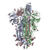
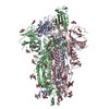
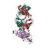
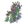
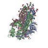
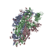
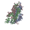
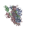
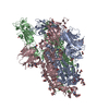
| Entry | Database: PDB / ID: 6zow | ||||||||||||||||||
|---|---|---|---|---|---|---|---|---|---|---|---|---|---|---|---|---|---|---|---|
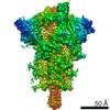
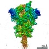
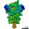
| Title | SARS-CoV-2 spike in prefusion state | ||||||||||||||||||
 Components Components | Spike glycoprotein | ||||||||||||||||||
 Keywords Keywords | VIRAL PROTEIN / Spike / Prefusion | ||||||||||||||||||
| Function / homology |  Function and homology information Function and homology informationsymbiont-mediated disruption of host tissue / Maturation of spike protein / Translation of Structural Proteins / Virion Assembly and Release / host cell surface / host extracellular region / symbiont-mediated-mediated suppression of host tetherin activity / Induction of Cell-Cell Fusion / structural constituent of virion / positive regulation of viral entry into host cell ...symbiont-mediated disruption of host tissue / Maturation of spike protein / Translation of Structural Proteins / Virion Assembly and Release / host cell surface / host extracellular region / symbiont-mediated-mediated suppression of host tetherin activity / Induction of Cell-Cell Fusion / structural constituent of virion / positive regulation of viral entry into host cell / membrane fusion / host cell endoplasmic reticulum-Golgi intermediate compartment membrane / Attachment and Entry / entry receptor-mediated virion attachment to host cell / receptor-mediated virion attachment to host cell / host cell surface receptor binding / symbiont-mediated suppression of host innate immune response / endocytosis involved in viral entry into host cell / receptor ligand activity / fusion of virus membrane with host plasma membrane / fusion of virus membrane with host endosome membrane / viral envelope / symbiont entry into host cell / virion attachment to host cell / host cell plasma membrane / SARS-CoV-2 activates/modulates innate and adaptive immune responses / virion membrane / membrane / identical protein binding / plasma membrane Similarity search - Function | ||||||||||||||||||
| Biological species |   Severe acute respiratory syndrome coronavirus 2 Severe acute respiratory syndrome coronavirus 2 | ||||||||||||||||||
| Method | ELECTRON MICROSCOPY / single particle reconstruction / cryo EM / Resolution: 3 Å | ||||||||||||||||||
 Authors Authors | Martinez, M. / Marabini, R. / Carazo, J.M. | ||||||||||||||||||
| Funding support |  Spain, European Union, Spain, European Union,  United States, 5items United States, 5items
| ||||||||||||||||||
 Citation Citation | Journal: IUCrJ / Year: 2020 Title: Continuous flexibility analysis of SARS-CoV-2 spike prefusion structures. Authors: Roberto Melero / Carlos Oscar S Sorzano / Brent Foster / José-Luis Vilas / Marta Martínez / Roberto Marabini / Erney Ramírez-Aportela / Ruben Sanchez-Garcia / David Herreros / Laura Del ...Authors: Roberto Melero / Carlos Oscar S Sorzano / Brent Foster / José-Luis Vilas / Marta Martínez / Roberto Marabini / Erney Ramírez-Aportela / Ruben Sanchez-Garcia / David Herreros / Laura Del Caño / Patricia Losana / Yunior C Fonseca-Reyna / Pablo Conesa / Daniel Wrapp / Pablo Chacon / Jason S McLellan / Hemant D Tagare / Jose-Maria Carazo / Abstract: Using a new consensus-based image-processing approach together with principal component analysis, the flexibility and conformational dynamics of the SARS-CoV-2 spike in the prefusion state have been ...Using a new consensus-based image-processing approach together with principal component analysis, the flexibility and conformational dynamics of the SARS-CoV-2 spike in the prefusion state have been analysed. These studies revealed concerted motions involving the receptor-binding domain (RBD), N-terminal domain, and subdomains 1 and 2 around the previously characterized 1-RBD-up state, which have been modeled as elastic deformations. It is shown that in this data set there are not well defined, stable spike conformations, but virtually a continuum of states. An ensemble map was obtained with minimum bias, from which the extremes of the change along the direction of maximal variance were modeled by flexible fitting. The results provide a warning of the potential image-processing classification instability of these complicated data sets, which has a direct impact on the interpretability of the results. #1: Journal: bioRxiv / Year: 2020 Title: Continuous flexibility analysis of SARS-CoV-2 Spike prefusion structures. Authors: Roberto Melero / Carlos Oscar S Sorzano / Brent Foster / José-Luis Vilas / Marta Martínez / Roberto Marabini / Erney Ramírez-Aportela / Ruben Sanchez-Garcia / David Herreros / Laura Del ...Authors: Roberto Melero / Carlos Oscar S Sorzano / Brent Foster / José-Luis Vilas / Marta Martínez / Roberto Marabini / Erney Ramírez-Aportela / Ruben Sanchez-Garcia / David Herreros / Laura Del Caño / Patricia Losana / Yunior C Fonseca-Reyna / Pablo Conesa / Daniel Wrapp / Pablo Chacon / Jason S McLellan / Hemant D Tagare / Jose-Maria Carazo / Abstract: With the help of novel processing workflows and algorithms, we have obtained a better understanding of the flexibility and conformational dynamics of the SARS-CoV-2 spike in the prefusion state. We ...With the help of novel processing workflows and algorithms, we have obtained a better understanding of the flexibility and conformational dynamics of the SARS-CoV-2 spike in the prefusion state. We have re-analyzed previous cryo-EM data combining 3D clustering approaches with ways to explore a continuous flexibility space based on 3D Principal Component Analysis. These advanced analyses revealed a concerted motion involving the receptor-binding domain (RBD), N-terminal domain (NTD), and subdomain 1 and 2 (SD1 & SD2) around the previously characterized 1-RBD-up state, which have been modeled as elastic deformations. We show that in this dataset there are not well-defined, stable, spike conformations, but virtually a continuum of states moving in a concerted fashion. We obtained an improved resolution ensemble map with minimum bias, from which we model by flexible fitting the extremes of the change along the direction of maximal variance. Moreover, a high-resolution structure of a recently described biochemically stabilized form of the spike is shown to greatly reduce the dynamics observed for the wild-type spike. Our results provide new detailed avenues to potentially restrain the spike dynamics for structure-based drug and vaccine design and at the same time give a warning of the potential image processing classification instability of these complicated datasets, having a direct impact on the interpretability of the results. | ||||||||||||||||||
| History |
|
- Structure visualization
| Movie |
Movie viewer |
|---|---|
| Structure viewer | Molecule: MolmilJmol/JSmol |
- Downloads & links
-Download
| PDBx/mmCIF format | 6zow.cif.gz | 564 KB | Display | PDBx/mmCIF format |
|---|---|---|---|---|
| PDB format | pdb6zow.ent.gz | 444 KB | Display | PDB format |
| PDBx/mmJSON format | 6zow.json.gz | Tree view | PDBx/mmJSON format | |
| Others |  Other downloads Other downloads |
-Validation report
| Arichive directory | https://data.pdbj.org/pub/pdb/validation_reports/zo/6zowftp://data.pdbj.org/pub/pdb/validation_reports/zo/6zow | HTTPS FTP |
|---|
-Related structure data
| Related structure data |  11328MC 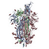 6zp5C 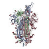 6zp7C M: map data used to model this data C: citing same article ( |
|---|---|
| Similar structure data | |
| EM raw data | EMPIAR-10516 (Title: Cryo electron microscopy of SARS-CoV-2 spike in prefusion state Data size: 2.1 TB / Data #1: 2.outputMovies [micrographs - multiframe]) |


-Links
 PDBj
PDBj


- Assembly
| Deposited unit | 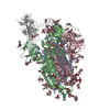
|
|---|---|
| 1 |
|
-Components
-Protein , 1 types, 4 molecules ACBa
| #1: Protein | Mass: 142399.375 Da / Num. of mol.: 4 / Mutation: K986P, E987P Source method: isolated from a genetically manipulated source Source: (gene. exp.) Severe acute respiratory syndrome coronavirus 2Gene: S, 2 / Plasmid: palphaH / Cell line (production host): FreeStyle293F / Production host:  Homo sapiens (human) / References: UniProt: P0DTC2 Homo sapiens (human) / References: UniProt: P0DTC2 |
|---|
-Sugars , 6 types, 47 molecules 

| #2: Polysaccharide | 2-acetamido-2-deoxy-beta-D-glucopyranose-(1-4)-2-acetamido-2-deoxy-beta-D-glucopyranose Source method: isolated from a genetically manipulated source #3: Polysaccharide | alpha-D-mannopyranose-(1-3)-beta-D-mannopyranose-(1-4)-2-acetamido-2-deoxy-beta-D-glucopyranose-(1- ...alpha-D-mannopyranose-(1-3)-beta-D-mannopyranose-(1-4)-2-acetamido-2-deoxy-beta-D-glucopyranose-(1-4)-2-acetamido-2-deoxy-beta-D-glucopyranose Source method: isolated from a genetically manipulated source #4: Polysaccharide | beta-D-mannopyranose-(1-4)-2-acetamido-2-deoxy-beta-D-glucopyranose-(1-4)-2-acetamido-2-deoxy-beta- ...beta-D-mannopyranose-(1-4)-2-acetamido-2-deoxy-beta-D-glucopyranose-(1-4)-2-acetamido-2-deoxy-beta-D-glucopyranose Source method: isolated from a genetically manipulated source #5: Polysaccharide | beta-D-mannopyranose-(1-4)-2-acetamido-2-deoxy-beta-D-glucopyranose-(1-6)-2-acetamido-2-deoxy-beta- ...beta-D-mannopyranose-(1-4)-2-acetamido-2-deoxy-beta-D-glucopyranose-(1-6)-2-acetamido-2-deoxy-beta-D-glucopyranose | Source method: isolated from a genetically manipulated source #6: Sugar | ChemComp-NAG /  Type: D-saccharide, beta linking / Mass: 221.208 Da / Num. of mol.: 18 Type: D-saccharide, beta linking / Mass: 221.208 Da / Num. of mol.: 18Source method: isolated from a genetically manipulated source Formula: C8H15NO6 #7: Sugar |  Type: D-saccharide, alpha linking / Mass: 180.156 Da / Num. of mol.: 2 Type: D-saccharide, alpha linking / Mass: 180.156 Da / Num. of mol.: 2Source method: isolated from a genetically manipulated source Formula: C6H12O6 |
|---|
-Non-polymers , 2 types, 7 molecules 

| #8: Chemical |  Mass: 78.133 Da / Num. of mol.: 3 / Source method: obtained synthetically / Formula: C2H6OS / Comment: DMSO, precipitant*YM Mass: 78.133 Da / Num. of mol.: 3 / Source method: obtained synthetically / Formula: C2H6OS / Comment: DMSO, precipitant*YM#9: Water | ChemComp-HOH / | Mass: 18.015 Da / Num. of mol.: 4 / Source method: isolated from a natural source / Formula: H2O |
|---|
-Details
| Has ligand of interest | N |
|---|---|
| Has protein modification | Y |
-Experimental details
-Experiment
| Experiment | Method: ELECTRON MICROSCOPY |
|---|---|
| EM experiment | Aggregation state: PARTICLE / 3D reconstruction method: single particle reconstruction |
- Sample preparation
| Component | Name: Spike homotrimer in prefusion state / Type: COMPLEX / Entity ID: #1 / Source: RECOMBINANT |
|---|---|
| Source (natural) | Organism: Severe acute respiratory syndrome coronavirus 2 |
| Source (recombinant) | Organism: Homo sapiens (human) / Cell: FreeStyle293F / Plasmid: palphaH |
| Buffer solution | pH: 8 |
| Specimen | Embedding applied: NO / Shadowing applied: NO / Staining applied: NO / Vitrification applied: YES |
| Vitrification | Cryogen name: ETHANE |
- Electron microscopy imaging
| Experimental equipment |  Model: Titan Krios / Image courtesy: FEI Company |
|---|---|
| Microscopy | Model: FEI TITAN KRIOS |
| Electron gun | Electron source:  FIELD EMISSION GUN / Accelerating voltage: 300 kV / Illumination mode: FLOOD BEAM FIELD EMISSION GUN / Accelerating voltage: 300 kV / Illumination mode: FLOOD BEAM |
| Electron lens | Mode: BRIGHT FIELD |
| Image recording | Electron dose: 36 e/Å2 / Film or detector model: GATAN K3 (6k x 4k) |
- Processing
| EM software |
| ||||||||||||||||||||||||||||||||||||||||||||||||||||||||||||
|---|---|---|---|---|---|---|---|---|---|---|---|---|---|---|---|---|---|---|---|---|---|---|---|---|---|---|---|---|---|---|---|---|---|---|---|---|---|---|---|---|---|---|---|---|---|---|---|---|---|---|---|---|---|---|---|---|---|---|---|---|---|
| CTF correction | Type: PHASE FLIPPING AND AMPLITUDE CORRECTION | ||||||||||||||||||||||||||||||||||||||||||||||||||||||||||||
| 3D reconstruction | Resolution: 3 Å / Resolution method: FSC 0.143 CUT-OFF / Num. of particles: 203000 / Symmetry type: POINT | ||||||||||||||||||||||||||||||||||||||||||||||||||||||||||||
| Atomic model building | Protocol: FLEXIBLE FIT / Space: REAL Details: Initial models 6VSB and 6VYB were used for flexible fitting, whereas 6YLA was used for rigid fitting of the RBD domain of chain A, that we have called a in the final structure. | ||||||||||||||||||||||||||||||||||||||||||||||||||||||||||||
| Atomic model building | 3D fitting-ID: 1 / Source name: PDB / Type: experimental model
|