Movie
Movie Controller

[English] 日本語
 Yorodumi
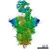
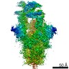
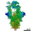
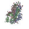
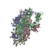
- PDB-7kj5: SARS-CoV-2 Spike Glycoprotein, prefusion with one RBD up conformation -
+ Open data

- Basic information
| Entry | Database: PDB / ID: 7kj5 | |||||||||||||||
|---|---|---|---|---|---|---|---|---|---|---|---|---|---|---|---|---|
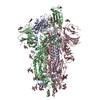
| Title | SARS-CoV-2 Spike Glycoprotein, prefusion with one RBD up conformation | |||||||||||||||
 Components Components | Spike glycoprotein | |||||||||||||||
 Keywords Keywords | VIRAL PROTEIN | |||||||||||||||
| Function / homology |  Function and homology information Function and homology informationsymbiont-mediated disruption of host tissue / Maturation of spike protein / Translation of Structural Proteins / Virion Assembly and Release / host cell surface / host extracellular region / symbiont-mediated-mediated suppression of host tetherin activity / Induction of Cell-Cell Fusion / structural constituent of virion / positive regulation of viral entry into host cell ...symbiont-mediated disruption of host tissue / Maturation of spike protein / Translation of Structural Proteins / Virion Assembly and Release / host cell surface / host extracellular region / symbiont-mediated-mediated suppression of host tetherin activity / Induction of Cell-Cell Fusion / structural constituent of virion / positive regulation of viral entry into host cell / membrane fusion / host cell endoplasmic reticulum-Golgi intermediate compartment membrane / Attachment and Entry / entry receptor-mediated virion attachment to host cell / receptor-mediated virion attachment to host cell / host cell surface receptor binding / symbiont-mediated suppression of host innate immune response / endocytosis involved in viral entry into host cell / receptor ligand activity / fusion of virus membrane with host plasma membrane / fusion of virus membrane with host endosome membrane / viral envelope / symbiont entry into host cell / virion attachment to host cell / host cell plasma membrane / SARS-CoV-2 activates/modulates innate and adaptive immune responses / virion membrane / membrane / identical protein binding / plasma membrane Similarity search - Function | |||||||||||||||
| Biological species |   Severe acute respiratory syndrome coronavirus 2 Severe acute respiratory syndrome coronavirus 2 | |||||||||||||||
| Method | ELECTRON MICROSCOPY / single particle reconstruction / cryo EM / Resolution: 3.6 Å | |||||||||||||||
 Authors Authors | Zhang, J. / Xiao, T.S. / Cai, Y.F. / Chen, B. | |||||||||||||||
| Funding support |  United States, 4items United States, 4items
| |||||||||||||||
 Citation Citation | Journal: Nat Struct Mol Biol / Year: 2021 Title: A trimeric human angiotensin-converting enzyme 2 as an anti-SARS-CoV-2 agent. Authors: Tianshu Xiao / Jianming Lu / Jun Zhang / Rebecca I Johnson / Lindsay G A McKay / Nadia Storm / Christy L Lavine / Hanqin Peng / Yongfei Cai / Sophia Rits-Volloch / Shen Lu / Brian D Quinlan ...Authors: Tianshu Xiao / Jianming Lu / Jun Zhang / Rebecca I Johnson / Lindsay G A McKay / Nadia Storm / Christy L Lavine / Hanqin Peng / Yongfei Cai / Sophia Rits-Volloch / Shen Lu / Brian D Quinlan / Michael Farzan / Michael S Seaman / Anthony Griffiths / Bing Chen / Abstract: Effective intervention strategies are urgently needed to control the COVID-19 pandemic. Human angiotensin-converting enzyme 2 (ACE2) is a membrane-bound carboxypeptidase that forms a dimer and serves ...Effective intervention strategies are urgently needed to control the COVID-19 pandemic. Human angiotensin-converting enzyme 2 (ACE2) is a membrane-bound carboxypeptidase that forms a dimer and serves as the cellular receptor for severe acute respiratory syndrome coronavirus 2 (SARS-CoV-2). ACE2 is also a key negative regulator of the renin-angiotensin system that modulates vascular functions. We report here the properties of a trimeric ACE2 ectodomain variant, engineered using a structure-based approach. The trimeric ACE2 variant has a binding affinity of ~60 pM for the spike protein of SARS‑CoV‑2 (compared with 77 nM for monomeric ACE2 and 12-22 nM for dimeric ACE2 constructs), and its peptidase activity and the ability to block activation of angiotensin II receptor type 1 in the renin-angiotensin system are preserved. Moreover, the engineered ACE2 potently inhibits SARS‑CoV‑2 infection in cell culture. These results suggest that engineered, trimeric ACE2 may be a promising anti-SARS-CoV-2 agent for treating COVID-19. #1: Journal: bioRxiv / Year: 2020 Title: A trimeric human angiotensin-converting enzyme 2 as an anti-SARS-CoV-2 agent in vitro. Authors: Tianshu Xiao / Jianming Lu / Jun Zhang / Rebecca I Johnson / Lindsay G A McKay / Nadia Storm / Christy L Lavine / Hanqin Peng / Yongfei Cai / Sophia Rits-Volloch / Shen Lu / Brian D Quinlan ...Authors: Tianshu Xiao / Jianming Lu / Jun Zhang / Rebecca I Johnson / Lindsay G A McKay / Nadia Storm / Christy L Lavine / Hanqin Peng / Yongfei Cai / Sophia Rits-Volloch / Shen Lu / Brian D Quinlan / Michael Farzan / Michael S Seaman / Anthony Griffiths / Bing Chen Abstract: Effective intervention strategies are urgently needed to control the COVID-19 pandemic. Human angiotensin-converting enzyme 2 (ACE2) is a carboxypeptidase that forms a dimer and serves as the ...Effective intervention strategies are urgently needed to control the COVID-19 pandemic. Human angiotensin-converting enzyme 2 (ACE2) is a carboxypeptidase that forms a dimer and serves as the cellular receptor for SARS-CoV-2. It is also a key negative regulator of the renin-angiotensin system (RAS), conserved in mammals, which modulates vascular functions. We report here the properties of a trimeric ACE2 variant, created by a structure-based approach, with binding affinity of ~60 pM for the spike (S) protein of SARS-CoV-2, while preserving the wildtype peptidase activity as well as the ability to block activation of angiotensin II receptor type 1 in the RAS. Moreover, the engineered ACE2 potently inhibits infection of SARS-CoV-2 in cell culture. These results suggest that engineered, trimeric ACE2 may be a promising anti-SARS-CoV-2 agent for treating COVID-19. | |||||||||||||||
| History |
|
- Structure visualization
| Movie |
Movie viewer |
|---|---|
| Structure viewer | Molecule: MolmilJmol/JSmol |
- Downloads & links
-Download
| PDBx/mmCIF format | 7kj5.cif.gz | 596.3 KB | Display | PDBx/mmCIF format |
|---|---|---|---|---|
| PDB format | pdb7kj5.ent.gz | 472.1 KB | Display | PDB format |
| PDBx/mmJSON format | 7kj5.json.gz | Tree view | PDBx/mmJSON format | |
| Others |  Other downloads Other downloads |
-Validation report
| Arichive directory | https://data.pdbj.org/pub/pdb/validation_reports/kj/7kj5ftp://data.pdbj.org/pub/pdb/validation_reports/kj/7kj5 | HTTPS FTP |
|---|
-Related structure data
| Related structure data | 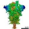 22894MC 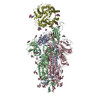 7kj2C 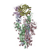 7kj3C 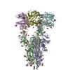 7kj4C M: map data used to model this data C: citing same article ( |
|---|---|
| Similar structure data |


-Links
 PDBj
PDBj


- Assembly
| Deposited unit | 
|
|---|---|
| 1 |
|
-Components
| #1: Protein | Mass: 136559.938 Da / Num. of mol.: 3 / Mutation: K986P,V987P Source method: isolated from a genetically manipulated source Source: (gene. exp.) Severe acute respiratory syndrome coronavirus 2Gene: S, 2 / Production host:  Homo sapiens (human) / References: UniProt: P0DTC2 Homo sapiens (human) / References: UniProt: P0DTC2#2: Polysaccharide | 2-acetamido-2-deoxy-beta-D-glucopyranose-(1-4)-2-acetamido-2-deoxy-beta-D-glucopyranose Source method: isolated from a genetically manipulated source #3: Sugar | ChemComp-NAG /   Type: D-saccharide, beta linking / Mass: 221.208 Da / Num. of mol.: 35 / Source method: obtained synthetically / Formula: C8H15NO6 / Feature type: SUBJECT OF INVESTIGATION Type: D-saccharide, beta linking / Mass: 221.208 Da / Num. of mol.: 35 / Source method: obtained synthetically / Formula: C8H15NO6 / Feature type: SUBJECT OF INVESTIGATIONHas ligand of interest | Y | Has protein modification | Y | |
|---|
-Experimental details
-Experiment
| Experiment | Method: ELECTRON MICROSCOPY |
|---|---|
| EM experiment | Aggregation state: PARTICLE / 3D reconstruction method: single particle reconstruction |
- Sample preparation
| Component | Name: SARS-CoV-2 Spike Glycoprotein, prefusion with one RBD-up conformation Type: COMPLEX Details: SARS-CoV-2 Spike Glycoprotein, prefusion with one RBD-up conformation Entity ID: #1 / Source: RECOMBINANT | |||||||||||||||
|---|---|---|---|---|---|---|---|---|---|---|---|---|---|---|---|---|
| Molecular weight | Value: 410 kDa/nm / Experimental value: NO | |||||||||||||||
| Source (natural) | Organism: Severe acute respiratory syndrome coronavirus 2 | |||||||||||||||
| Source (recombinant) | Organism: Homo sapiens (human) | |||||||||||||||
| Buffer solution | pH: 7.5 | |||||||||||||||
| Buffer component |
| |||||||||||||||
| Specimen | Conc.: 1 mg/ml / Embedding applied: NO / Shadowing applied: NO / Staining applied: NO / Vitrification applied: YES | |||||||||||||||
| Vitrification | Instrument: FEI VITROBOT MARK IV / Cryogen name: ETHANE / Humidity: 100 % / Chamber temperature: 277.15 K |
- Electron microscopy imaging
| Experimental equipment |  Model: Titan Krios / Image courtesy: FEI Company |
|---|---|
| Microscopy | Model: FEI TITAN KRIOS |
| Electron gun | Electron source:  FIELD EMISSION GUN / Accelerating voltage: 300 kV / Illumination mode: FLOOD BEAM FIELD EMISSION GUN / Accelerating voltage: 300 kV / Illumination mode: FLOOD BEAM |
| Electron lens | Mode: BRIGHT FIELD |
| Image recording | Electron dose: 50.05 e/Å2 / Film or detector model: GATAN K3 BIOQUANTUM (6k x 4k) |
- Processing
| Software | Name: PHENIX / Version: 1.18.2_3874: / Classification: refinement | ||||||||||||||||||||||||
|---|---|---|---|---|---|---|---|---|---|---|---|---|---|---|---|---|---|---|---|---|---|---|---|---|---|
| EM software |
| ||||||||||||||||||||||||
| CTF correction | Type: PHASE FLIPPING AND AMPLITUDE CORRECTION | ||||||||||||||||||||||||
| Particle selection | Num. of particles selected: 407761 | ||||||||||||||||||||||||
| 3D reconstruction | Resolution: 3.6 Å / Resolution method: FSC 0.143 CUT-OFF / Num. of particles: 32685 / Symmetry type: POINT | ||||||||||||||||||||||||
| Atomic model building | Protocol: AB INITIO MODEL | ||||||||||||||||||||||||
| Atomic model building | PDB-ID: 6VYB Pdb chain-ID: A / Accession code: 6VYB / Pdb chain residue range: 14-1211 / Source name: PDB / Type: experimental model | ||||||||||||||||||||||||
| Refine LS restraints |
|