 Movie
Movie Controller


+ Open data

- Basic information
| Entry | Database: PDB / ID: 6x2b | ||||||||||||||||||
|---|---|---|---|---|---|---|---|---|---|---|---|---|---|---|---|---|---|---|---|
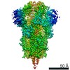
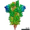
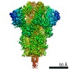
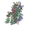
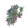
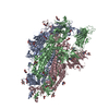
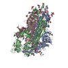
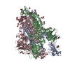
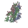
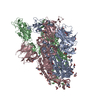
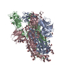
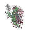
| Title | SARS-CoV-2 u1S2q 2-RBD Up Spike Protein Trimer | ||||||||||||||||||
 Components Components | Spike glycoprotein | ||||||||||||||||||
 Keywords Keywords | VIRAL PROTEIN / Trimer | ||||||||||||||||||
| Function / homology |  Function and homology information Function and homology informationsymbiont-mediated disruption of host tissue / Maturation of spike protein / Translation of Structural Proteins / Virion Assembly and Release / host cell surface / host extracellular region / symbiont-mediated-mediated suppression of host tetherin activity / Induction of Cell-Cell Fusion / structural constituent of virion / positive regulation of viral entry into host cell ...symbiont-mediated disruption of host tissue / Maturation of spike protein / Translation of Structural Proteins / Virion Assembly and Release / host cell surface / host extracellular region / symbiont-mediated-mediated suppression of host tetherin activity / Induction of Cell-Cell Fusion / structural constituent of virion / positive regulation of viral entry into host cell / membrane fusion / host cell endoplasmic reticulum-Golgi intermediate compartment membrane / Attachment and Entry / entry receptor-mediated virion attachment to host cell / receptor-mediated virion attachment to host cell / host cell surface receptor binding / symbiont-mediated suppression of host innate immune response / endocytosis involved in viral entry into host cell / receptor ligand activity / fusion of virus membrane with host plasma membrane / fusion of virus membrane with host endosome membrane / viral envelope / symbiont entry into host cell / virion attachment to host cell / host cell plasma membrane / SARS-CoV-2 activates/modulates innate and adaptive immune responses / virion membrane / membrane / identical protein binding / plasma membrane Similarity search - Function | ||||||||||||||||||
| Biological species |   Severe acute respiratory syndrome coronavirus 2 Severe acute respiratory syndrome coronavirus 2 | ||||||||||||||||||
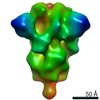
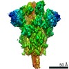
| Method | ELECTRON MICROSCOPY / single particle reconstruction / cryo EM / Resolution: 3.6 Å | ||||||||||||||||||
 Authors Authors | Henderson, R. / Acharya, P. | ||||||||||||||||||
| Funding support |  United States, 5items United States, 5items
| ||||||||||||||||||
 Citation Citation | Journal: Nat Struct Mol Biol / Year: 2020 Title: Controlling the SARS-CoV-2 spike glycoprotein conformation. Authors: Rory Henderson / Robert J Edwards / Katayoun Mansouri / Katarzyna Janowska / Victoria Stalls / Sophie M C Gobeil / Megan Kopp / Dapeng Li / Rob Parks / Allen L Hsu / Mario J Borgnia / Barton ...Authors: Rory Henderson / Robert J Edwards / Katayoun Mansouri / Katarzyna Janowska / Victoria Stalls / Sophie M C Gobeil / Megan Kopp / Dapeng Li / Rob Parks / Allen L Hsu / Mario J Borgnia / Barton F Haynes / Priyamvada Acharya / Abstract: The coronavirus (CoV) spike (S) protein, involved in viral-host cell fusion, is the primary immunogenic target for virus neutralization and the current focus of many vaccine design efforts. The ...The coronavirus (CoV) spike (S) protein, involved in viral-host cell fusion, is the primary immunogenic target for virus neutralization and the current focus of many vaccine design efforts. The highly flexible S-protein, with its mobile domains, presents a moving target to the immune system. Here, to better understand S-protein mobility, we implemented a structure-based vector analysis of available β-CoV S-protein structures. Despite an overall similarity in domain organization, we found that S-proteins from different β-CoVs display distinct configurations. Based on this analysis, we developed two soluble ectodomain constructs for the SARS-CoV-2 S-protein, in which the highly immunogenic and mobile receptor binding domain (RBD) is either locked in the all-RBDs 'down' position or adopts 'up' state conformations more readily than the wild-type S-protein. These results demonstrate that the conformation of the S-protein can be controlled via rational design and can provide a framework for the development of engineered CoV S-proteins for vaccine applications. #1: Journal: bioRxiv / Year: 2020 Title: Controlling the SARS-CoV-2 Spike Glycoprotein Conformation. Authors: Rory Henderson / Robert J Edwards / Katayoun Mansouri / Katarzyna Janowska / Victoria Stalls / Sophie Gobeil / Megan Kopp / Allen Hsu / Mario Borgnia / Rob Parks / Barton F Haynes / Priyamvada Acharya / Abstract: The coronavirus (CoV) viral host cell fusion spike (S) protein is the primary immunogenic target for virus neutralization and the current focus of many vaccine design efforts. The highly flexible S- ...The coronavirus (CoV) viral host cell fusion spike (S) protein is the primary immunogenic target for virus neutralization and the current focus of many vaccine design efforts. The highly flexible S-protein, with its mobile domains, presents a moving target to the immune system. Here, to better understand S-protein mobility, we implemented a structure-based vector analysis of available β-CoV S-protein structures. We found that despite overall similarity in domain organization, different β-CoV strains display distinct S-protein configurations. Based on this analysis, we developed two soluble ectodomain constructs in which the highly immunogenic and mobile receptor binding domain (RBD) is locked in either the all-RBDs 'down' position or is induced to display a previously unobserved in SARS-CoV-2 2-RBDs 'up' configuration. These results demonstrate that the conformation of the S-protein can be controlled via rational design and provide a framework for the development of engineered coronavirus spike proteins for vaccine applications. | ||||||||||||||||||
| History |
|
- Structure visualization
| Movie |
Movie viewer |
|---|---|
| Structure viewer | Molecule: MolmilJmol/JSmol |
- Downloads & links
-Download
| PDBx/mmCIF format | 6x2b.cif.gz | 508.6 KB | Display | PDBx/mmCIF format |
|---|---|---|---|---|
| PDB format | pdb6x2b.ent.gz | 390.1 KB | Display | PDB format |
| PDBx/mmJSON format | 6x2b.json.gz | Tree view | PDBx/mmJSON format | |
| Others |  Other downloads Other downloads |
-Validation report
| Arichive directory | https://data.pdbj.org/pub/pdb/validation_reports/x2/6x2bftp://data.pdbj.org/pub/pdb/validation_reports/x2/6x2b | HTTPS FTP |
|---|
-Related structure data
| Related structure data | 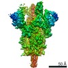 22000MC 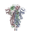 6x29C 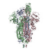 6x2aC 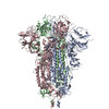 6x2cC M: map data used to model this data C: citing same article ( |
|---|---|
| Similar structure data |
-Links
 PDBj
PDBj


- Assembly
| Deposited unit | 
|
|---|---|
| 1 |
|
-Components
| #1: Protein | Mass: 140790.453 Da / Num. of mol.: 3 / Fragment: ectodomain (UNP residues 16-1208) / Mutation: F855Y+N856I+A570L+T572I Source method: isolated from a genetically manipulated source Source: (gene. exp.) Severe acute respiratory syndrome coronavirus 2Gene: S, 2 / Production host:  Homo sapiens (human) / References: UniProt: P0DTC2 Homo sapiens (human) / References: UniProt: P0DTC2Has protein modification | Y | |
|---|
-Experimental details
-Experiment
| Experiment | Method: ELECTRON MICROSCOPY |
|---|---|
| EM experiment | Aggregation state: PARTICLE / 3D reconstruction method: single particle reconstruction |
- Sample preparation
| Component | Name: Severe acute respiratory syndrome coronavirus 2 / Type: VIRUS / Entity ID: all / Source: RECOMBINANT |
|---|---|
| Source (natural) | Organism: Severe acute respiratory syndrome coronavirus 2 |
| Source (recombinant) | Organism: Homo sapiens (human) |
| Details of virus | Empty: NO / Enveloped: YES / Isolate: OTHER / Type: VIRION |
| Buffer solution | pH: 7.4 |
| Specimen | Embedding applied: NO / Shadowing applied: NO / Staining applied: NO / Vitrification applied: YES |
| Specimen support | Details: unspecified |
| Vitrification | Cryogen name: ETHANE |
- Electron microscopy imaging
| Experimental equipment |  Model: Titan Krios / Image courtesy: FEI Company |
|---|---|
| Microscopy | Model: FEI TITAN KRIOS |
| Electron gun | Electron source:  FIELD EMISSION GUN / Accelerating voltage: 300 kV / Illumination mode: FLOOD BEAM FIELD EMISSION GUN / Accelerating voltage: 300 kV / Illumination mode: FLOOD BEAM |
| Electron lens | Mode: BRIGHT FIELD |
| Image recording | Electron dose: 66.82 e/Å2 / Film or detector model: GATAN K3 (6k x 4k) |
- Processing
| CTF correction | Type: PHASE FLIPPING AND AMPLITUDE CORRECTION |
|---|---|
| 3D reconstruction | Resolution: 3.6 Å / Resolution method: FSC 0.143 CUT-OFF / Num. of particles: 133957 / Symmetry type: POINT |
