Movie
Movie Controller


+ Open data

- Basic information
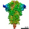
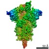
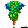
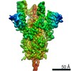
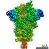
| Entry | Database: EMDB / ID: EMD-22809 | |||||||||
|---|---|---|---|---|---|---|---|---|---|---|
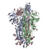
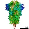
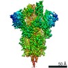
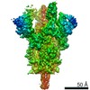
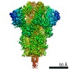
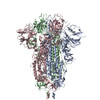
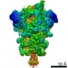
| Title | SARS-CoV-2 rS2d RBD-Down State Spike Protein Trimer | |||||||||
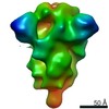
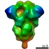
 Map data Map data | Sharpened and filtered map | |||||||||
 Sample Sample |
| |||||||||
| Biological species |   Severe acute respiratory syndrome coronavirus 2 Severe acute respiratory syndrome coronavirus 2 | |||||||||
| Method | single particle reconstruction / negative staining / Resolution: 12.06 Å | |||||||||
 Authors Authors | Edwards RJ / Mansouri K | |||||||||
| Funding support |  United States, 1 items United States, 1 items
| |||||||||
 Citation Citation | Journal: Nat Struct Mol Biol / Year: 2020 Title: Controlling the SARS-CoV-2 spike glycoprotein conformation. Authors: Rory Henderson / Robert J Edwards / Katayoun Mansouri / Katarzyna Janowska / Victoria Stalls / Sophie M C Gobeil / Megan Kopp / Dapeng Li / Rob Parks / Allen L Hsu / Mario J Borgnia / Barton ...Authors: Rory Henderson / Robert J Edwards / Katayoun Mansouri / Katarzyna Janowska / Victoria Stalls / Sophie M C Gobeil / Megan Kopp / Dapeng Li / Rob Parks / Allen L Hsu / Mario J Borgnia / Barton F Haynes / Priyamvada Acharya / Abstract: The coronavirus (CoV) spike (S) protein, involved in viral-host cell fusion, is the primary immunogenic target for virus neutralization and the current focus of many vaccine design efforts. The ...The coronavirus (CoV) spike (S) protein, involved in viral-host cell fusion, is the primary immunogenic target for virus neutralization and the current focus of many vaccine design efforts. The highly flexible S-protein, with its mobile domains, presents a moving target to the immune system. Here, to better understand S-protein mobility, we implemented a structure-based vector analysis of available β-CoV S-protein structures. Despite an overall similarity in domain organization, we found that S-proteins from different β-CoVs display distinct configurations. Based on this analysis, we developed two soluble ectodomain constructs for the SARS-CoV-2 S-protein, in which the highly immunogenic and mobile receptor binding domain (RBD) is either locked in the all-RBDs 'down' position or adopts 'up' state conformations more readily than the wild-type S-protein. These results demonstrate that the conformation of the S-protein can be controlled via rational design and can provide a framework for the development of engineered CoV S-proteins for vaccine applications. | |||||||||
| History |
|
- Structure visualization
| Movie |
 Movie viewer Movie viewer |
|---|---|
| Structure viewer | EM map: SurfViewMolmilJmol/JSmol |
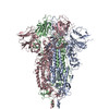
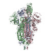
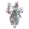
| Supplemental images |
 UCSF Chimera
- Downloads & links
-EMDB archive
| Map data | emd_22809.map.gz | 3.1 MB | EMDB map data format | |
|---|---|---|---|---|
| Header (meta data) | emd-22809-v30.xmlemd-22809.xml | 23.2 KB 23.2 KB | Display Display | EMDB header |
| FSC (resolution estimation) | emd_22809_fsc.xml | 3.6 KB | Display | FSC data file |
| Images |  emd_22809.png emd_22809.png | 41.3 KB | ||
| Masks | emd_22809_msk_1.map | 3.4 MB | Mask map | |
| Others | emd_22809_additional_1.map.gzemd_22809_half_map_1.map.gzemd_22809_half_map_2.map.gz | 2.4 MB 2.4 MB 2.4 MB | ||
| Archive directory |  http://ftp.pdbj.org/pub/emdb/structures/EMD-22809ftp://ftp.pdbj.org/pub/emdb/structures/EMD-22809 http://ftp.pdbj.org/pub/emdb/structures/EMD-22809ftp://ftp.pdbj.org/pub/emdb/structures/EMD-22809 | HTTPS FTP |
-Related structure data


-Links
| EMDB pages | EMDB (EBI/PDBe) / EMDataResource |
|---|
-Map
| File | Download / File: emd_22809.map.gz / Format: CCP4 / Size: 3.4 MB / Type: IMAGE STORED AS FLOATING POINT NUMBER (4 BYTES) | ||||||||||||||||||||||||||||||||||||||||||||||||||||||||||||||||||||
|---|---|---|---|---|---|---|---|---|---|---|---|---|---|---|---|---|---|---|---|---|---|---|---|---|---|---|---|---|---|---|---|---|---|---|---|---|---|---|---|---|---|---|---|---|---|---|---|---|---|---|---|---|---|---|---|---|---|---|---|---|---|---|---|---|---|---|---|---|---|
| Annotation | Sharpened and filtered map | ||||||||||||||||||||||||||||||||||||||||||||||||||||||||||||||||||||
| Projections & slices | Image control
Images are generated by Spider. | ||||||||||||||||||||||||||||||||||||||||||||||||||||||||||||||||||||
| Voxel size | X=Y=Z: 4.02 Å | ||||||||||||||||||||||||||||||||||||||||||||||||||||||||||||||||||||
| Density |
| ||||||||||||||||||||||||||||||||||||||||||||||||||||||||||||||||||||
| Symmetry | Space group: 1 | ||||||||||||||||||||||||||||||||||||||||||||||||||||||||||||||||||||
| Details | EMDB XML:
CCP4 map header:
| ||||||||||||||||||||||||||||||||||||||||||||||||||||||||||||||||||||
 Z (Sec.)
 Y (Row.)
 X (Col.)


-Supplemental data
-Mask #1
| File | emd_22809_msk_1.map | ||||||||||||
|---|---|---|---|---|---|---|---|---|---|---|---|---|---|
| Projections & Slices |
| ||||||||||||
| Density Histograms |


-Additional map: Unsharpened map
| File | emd_22809_additional_1.map | ||||||||||||
|---|---|---|---|---|---|---|---|---|---|---|---|---|---|
| Annotation | Unsharpened map | ||||||||||||
| Projections & Slices |
| ||||||||||||
| Density Histograms |


-Half map: half-map 1
| File | emd_22809_half_map_1.map | ||||||||||||
|---|---|---|---|---|---|---|---|---|---|---|---|---|---|
| Annotation | half-map 1 | ||||||||||||
| Projections & Slices |
| ||||||||||||
| Density Histograms |


-Half map: half-map 2
| File | emd_22809_half_map_2.map | ||||||||||||
|---|---|---|---|---|---|---|---|---|---|---|---|---|---|
| Annotation | half-map 2 | ||||||||||||
| Projections & Slices |
| ||||||||||||
| Density Histograms |


- Sample components
-Entire : SARS-CoV-2 rS2d Down State Spike Protein Trimer
| Entire | Name: SARS-CoV-2 rS2d Down State Spike Protein Trimer |
|---|---|
| Components |
|
-Supramolecule #1: SARS-CoV-2 rS2d Down State Spike Protein Trimer
| Supramolecule | Name: SARS-CoV-2 rS2d Down State Spike Protein Trimer / type: complex / ID: 1 / Parent: 0 / Macromolecule list: all |
|---|---|
| Source (natural) | Organism: Severe acute respiratory syndrome coronavirus 2 |
| Recombinant expression | Organism:  Homo sapiens (human) / Recombinant cell: 293F / Recombinant plasmid: p-alpha-H Homo sapiens (human) / Recombinant cell: 293F / Recombinant plasmid: p-alpha-H |
| Molecular weight | Theoretical: 481.8 KDa |
-Macromolecule #1: SARS-CoV-2 rS2d Down State Spike Protein Trimer
| Macromolecule | Name: SARS-CoV-2 rS2d Down State Spike Protein Trimer / type: protein_or_peptide / ID: 1 / Enantiomer: LEVO |
|---|---|
| Source (natural) | Organism: Severe acute respiratory syndrome coronavirus 2 |
| Recombinant expression | Organism: Homo sapiens (human) |
| Sequence | String: VNLTTRTQLP PAYTNSFTRG VYYPDKVFRS SVLHSTQDLF LPFFSNVTWF HAIHVSGTNG TKRFDNPVLP FNDGVYFAST EKSNIIRGWI FGTTLDSKTQ SLLIVNNATN VVIKVCEFQF CNDPFLGVYY HKNNKSWMES EFRVYSSANN CTFEYVSQPF LMDLEGKQGN ...String: VNLTTRTQLP PAYTNSFTRG VYYPDKVFRS SVLHSTQDLF LPFFSNVTWF HAIHVSGTNG TKRFDNPVLP FNDGVYFAST EKSNIIRGWI FGTTLDSKTQ SLLIVNNATN VVIKVCEFQF CNDPFLGVYY HKNNKSWMES EFRVYSSANN CTFEYVSQPF LMDLEGKQGN FKNLREFVFK NIDGYFKIYS KHTPINLVRD LPQGFSALEP LVDLPIGINI TRFQTLLALH RSYLTPGDSS SGWTAGAAAY YVGYLQPRTF LLKYNENGTI TDAVDCALDP LSETKCTLKS FTVEKGIYQT SNFRVQPTES IVRFPNITNL CPFGEVFNAT RFASVYAWNR KRISNCVADY SVLYNSASFS TFKCYGVCPT KLNDLCFTNV YADSFVIRGD EVRQIAPGQT GKIADYNYKL PDDFTGCVIA WNSNNLDSKV GGNYNYLYRL FRKSNLKPFE RDISTEIYQA GSTPCNGVEG FNCYFPLQSY GFQPTNGVGY QPYRVVVLSF ELLHAPATVC GPKKSTNLVK NKCVNFNFNG LTGTGVLTES NKKFLPFQQF GRDIADTTDA VRDPQTLEIL DITPCSFGGV SVITPGTNTS NQVAVLYQDV NCTEVPVAIH ADQLTPTWRV YSTGSNVFQT RAGCLIGAEH VNNSYECDIP IGAGICASYQ TQTNSPGSAS SVASQSIIAY TMSLGAENSV AYSNNSIAIP TNFTISVTTE ILPVSMTKTS VDCTMYICGD STECSNLLLQ YGSFCTQLNR ALTGIAVEQD KNTQEVFAQV KQIYKTPPIK DFGGFNFSQI LPDPSKPSKR SFIEDLLFNK VTLADAGFIK QYGDCLGDIA ARDLICAQKF NGLTVLPPLL TDEMIAQYTS ALLAGTITSG WTFGAGAALQ IPFAMQMAYR FNGIGVTQNV LYENQKLIAN QFNSAIGKIQ DSLSSTASAL GKLQDVVNQN AQALNTLVKQ LSSNFGAISS VLNDILSRLC PPEAEVQIDR LITGRLQSLQ TYVTQQLIRA AEIRASANLA ATKMSECVLG QSKRVDFCGK GYHLMSFPQS APHGVVFLHV TYVPAQEKNF TTAPAICHDG KAHFPREGVF VSNGTHWFVT QRNFYEPQII TTDNTFVSGN CDVVIGIVNN TVYDPLQPEL DSFKEELDKY FKNHTSPDVD LGDISGINAS VVNIQKEIDR LNEVAKNLNE SLIDLQELGK YEQGSGYIPE APRDGQAYVR KDGEWVLLST FLGRSLEVLF QGPGHHHHHH HHSAWSHPQF EKGGGSGGGG SGGSAWSHPQ FEK |
-Experimental details
-Structure determination
| Method | negative staining |
|---|---|
 Processing Processing | single particle reconstruction |
| Aggregation state | particle |
-Sample preparation
| Concentration | 0.1 mg/mL | ||||||||||||
|---|---|---|---|---|---|---|---|---|---|---|---|---|---|
| Buffer | pH: 7.4 Component:
| ||||||||||||
| Staining | Type: NEGATIVE / Material: Uranyl Formate Details: Samples were diluted to 0.1 mg/mL in 20 mM HEPES buffer, pH 7.4, with 5% glycerol, 150 mM NaCl, and 7.5 mM glutaraldehyde. After 5 minute incubation at room temperature, sufficient 1 M Tris ...Details: Samples were diluted to 0.1 mg/mL in 20 mM HEPES buffer, pH 7.4, with 5% glycerol, 150 mM NaCl, and 7.5 mM glutaraldehyde. After 5 minute incubation at room temperature, sufficient 1 M Tris stock, pH 7.4, was added to a final concentration of 75 mM Tris to quench unreacted glutaraldehyde, and was then incubated 5 minutes. Sample was then applied to a carbon film over 400 mesh copper EM grids that had been glow-discharged, incubated 1 minute, and then stained with 2% uranyl formate. | ||||||||||||
| Grid | Model: Homemade / Material: COPPER / Mesh: 400 / Support film - Material: CARBON / Support film - topology: CONTINUOUS / Support film - Film thickness: 0.05 nm / Pretreatment - Type: GLOW DISCHARGE |
- Electron microscopy
| Microscope | FEI/PHILIPS EM420 |
|---|---|
| Image recording | Film or detector model: OTHER / Digitization - Dimensions - Width: 2048 pixel / Digitization - Dimensions - Height: 2048 pixel / Digitization - Sampling interval: 33.0 µm / Number grids imaged: 1 / Number real images: 437 / Average exposure time: 0.5 sec. / Average electron dose: 32.0 e/Å2 |
| Electron beam | Acceleration voltage: 120 kV / Electron source: LAB6 |
| Electron optics | Illumination mode: FLOOD BEAM / Imaging mode: BRIGHT FIELD / Cs: 2.0 mm / Nominal defocus max: 1.5 µm / Nominal defocus min: 0.4 µm / Nominal magnification: 82000 |
| Sample stage | Specimen holder model: SIDE ENTRY, EUCENTRIC |