 Movie
Movie Controller


+ Open data

- Basic information
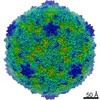
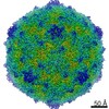
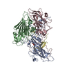
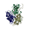
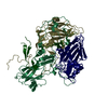
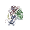
| Entry | Database: PDB / ID: 6zms | |||||||||
|---|---|---|---|---|---|---|---|---|---|---|
| Title | Coxsackievirus B4 strain E2 | |||||||||
 Components Components |
| |||||||||
 Keywords Keywords | VIRUS / Coxsackievirus B4 | |||||||||
| Function / homology |  Function and homology information Function and homology informationsymbiont-mediated suppression of host cytoplasmic pattern recognition receptor signaling pathway via inhibition of RIG-I activity / picornain 2A / symbiont-mediated suppression of host mRNA export from nucleus / symbiont genome entry into host cell via pore formation in plasma membrane / picornain 3C / T=pseudo3 icosahedral viral capsid / host cell cytoplasmic vesicle membrane / ribonucleoside triphosphate phosphatase activity / nucleoside-triphosphate phosphatase / channel activity ...symbiont-mediated suppression of host cytoplasmic pattern recognition receptor signaling pathway via inhibition of RIG-I activity / picornain 2A / symbiont-mediated suppression of host mRNA export from nucleus / symbiont genome entry into host cell via pore formation in plasma membrane / picornain 3C / T=pseudo3 icosahedral viral capsid / host cell cytoplasmic vesicle membrane / ribonucleoside triphosphate phosphatase activity / nucleoside-triphosphate phosphatase / channel activity / monoatomic ion transmembrane transport / DNA replication / RNA helicase activity / endocytosis involved in viral entry into host cell / symbiont-mediated activation of host autophagy / RNA-directed RNA polymerase / cysteine-type endopeptidase activity / viral RNA genome replication / RNA-directed RNA polymerase activity / virion attachment to host cell / host cell nucleus / structural molecule activity / DNA-templated transcription / proteolysis / RNA binding / zinc ion binding / ATP binding Similarity search - Function | |||||||||
| Biological species |  Coxsackievirus B4 Coxsackievirus B4 | |||||||||
| Method | ELECTRON MICROSCOPY / single particle reconstruction / cryo EM / Resolution: 3.4 Å | |||||||||
 Authors Authors | Flatt, J.W. / Domanska, A. / Butcher, S.J. | |||||||||
| Funding support |  Finland, 2items Finland, 2items
| |||||||||
 Citation Citation | Journal: Commun Biol / Year: 2021 Title: Identification of a conserved virion-stabilizing network inside the interprotomer pocket of enteroviruses. Authors: Justin W Flatt / Aušra Domanska / Alma L Seppälä / Sarah J Butcher / Abstract: Enteroviruses pose a persistent and widespread threat to human physical health, with no specific treatments available. Small molecule capsid binders have the potential to be developed as antivirals ...Enteroviruses pose a persistent and widespread threat to human physical health, with no specific treatments available. Small molecule capsid binders have the potential to be developed as antivirals that prevent virus attachment and entry into host cells. To aid with broad-range drug development, we report here structures of coxsackieviruses B3 and B4 bound to different interprotomer-targeting capsid binders using single-particle cryo-EM. The EM density maps are beyond 3 Å resolution, providing detailed information about interactions in the ligand-binding pocket. Comparative analysis revealed the residues that form a conserved virion-stabilizing network at the interprotomer site, and showed the small molecule properties that allow anchoring in the pocket to inhibit virus disassembly. | |||||||||
| History |
|
- Structure visualization
| Movie |
Movie viewer |
|---|---|
| Structure viewer | Molecule: MolmilJmol/JSmol |
- Downloads & links
-Download
| PDBx/mmCIF format | 6zms.cif.gz | 271.2 KB | Display | PDBx/mmCIF format |
|---|---|---|---|---|
| PDB format | pdb6zms.ent.gz | 215.5 KB | Display | PDB format |
| PDBx/mmJSON format | 6zms.json.gz | Tree view | PDBx/mmJSON format | |
| Others |  Other downloads Other downloads |
-Validation report
| Arichive directory | https://data.pdbj.org/pub/pdb/validation_reports/zm/6zmsftp://data.pdbj.org/pub/pdb/validation_reports/zm/6zms | HTTPS FTP |
|---|
-Related structure data
| Related structure data | 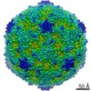 11300MC  6zckC  6zclC M: map data used to model this data C: citing same article ( |
|---|---|
| Similar structure data | |
| EM raw data | EMPIAR-10652 (Title: Identification of a conserved virion-stabilizing network inside the interprotomer pocket of enteroviruses Data size: 7.3 TB Data #1: Unaligned multiframe micrographs of CVB4 in complex with CP48 [micrographs - multiframe] Data #2: Aligned multi-frame micrographs dose weighted [micrographs - single frame] Data #3: Aligned non dose weighted micrographs [micrographs - single frame] Data #4: Un-aligned multiframe micrographs of CVB4 control [micrographs - multiframe]) |


-Links
 PDBj
PDBj

- Assembly
| Deposited unit | 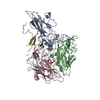
|
|---|---|
| 1 | x 60
|
-Components
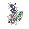
| #1: Protein | Mass: 30685.498 Da / Num. of mol.: 1 / Source method: isolated from a natural source / Source: (natural) Coxsackievirus B4 (strain E2) / Cell line: BGM / Strain: E2 / References: UniProt: Q86887 |
|---|---|
| #2: Protein | Mass: 27708.070 Da / Num. of mol.: 1 / Source method: isolated from a natural source / Source: (natural) Coxsackievirus B4 (strain E2) / Cell line: BGM / Strain: E2 / References: UniProt: Q86887 |
| #3: Protein | Mass: 26444.160 Da / Num. of mol.: 1 / Source method: isolated from a natural source / Source: (natural) Coxsackievirus B4 (strain E2) / Strain: E2 / References: UniProt: Q86887 |
| #4: Protein | Mass: 7499.235 Da / Num. of mol.: 1 / Source method: isolated from a natural source / Source: (natural) Coxsackievirus B4 (strain E2) / Cell line: BGM / Strain: E2 / References: UniProt: Q86887 |
| Has ligand of interest | N |
-Experimental details
-Experiment
| Experiment | Method: ELECTRON MICROSCOPY |
|---|---|
| EM experiment | Aggregation state: PARTICLE / 3D reconstruction method: single particle reconstruction |
- Sample preparation
| Component | Name: Coxsackievirus B4 (strain E2) / Type: VIRUS / Entity ID: all / Source: NATURAL | |||||||||||||||||||||||||
|---|---|---|---|---|---|---|---|---|---|---|---|---|---|---|---|---|---|---|---|---|---|---|---|---|---|---|
| Molecular weight | Experimental value: NO | |||||||||||||||||||||||||
| Source (natural) | Organism: Coxsackievirus B4 (strain E2) | |||||||||||||||||||||||||
| Details of virus | Empty: NO / Enveloped: NO / Isolate: STRAIN / Type: VIRION | |||||||||||||||||||||||||
| Natural host | Organism: Homo sapiens | |||||||||||||||||||||||||
| Virus shell | Name: icosahedral / Diameter: 300 nm / Triangulation number (T number): 3 | |||||||||||||||||||||||||
| Buffer solution | pH: 7 | |||||||||||||||||||||||||
| Buffer component |
| |||||||||||||||||||||||||
| Specimen | Embedding applied: NO / Shadowing applied: NO / Staining applied: NO / Vitrification applied: YES | |||||||||||||||||||||||||
| Vitrification | Cryogen name: ETHANE |
- Electron microscopy imaging
| Experimental equipment |  Model: Tecnai F20 / Image courtesy: FEI Company |
|---|---|
| Microscopy | Model: TFS TALOS F200C |
| Electron gun | Electron source:  FIELD EMISSION GUN / Accelerating voltage: 200 kV / Illumination mode: FLOOD BEAM FIELD EMISSION GUN / Accelerating voltage: 200 kV / Illumination mode: FLOOD BEAM |
| Electron lens | Mode: BRIGHT FIELD |
| Image recording | Electron dose: 40 e/Å2 / Film or detector model: FEI FALCON III (4k x 4k) |
- Processing
| CTF correction | Type: PHASE FLIPPING AND AMPLITUDE CORRECTION |
|---|---|
| 3D reconstruction | Resolution: 3.4 Å / Resolution method: FSC 0.143 CUT-OFF / Num. of particles: 40627 / Symmetry type: POINT |
| Refinement | Highest resolution: 3.4 Å |
