 Movie
Movie Controller


+ Open data

- Basic information
| Entry | Database: EMDB / ID: EMD-11166 | |||||||||
|---|---|---|---|---|---|---|---|---|---|---|
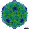
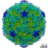
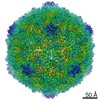
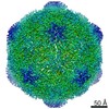
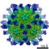
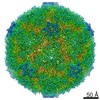
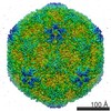
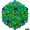
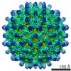
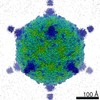
| Title | Coxsackievirus B3 in complex with capsid binder compound 17 | |||||||||
 Map data Map data | Coxsackie B3 virion in complex with compound 17 | |||||||||
 Sample Sample |
| |||||||||
 Keywords Keywords | Enterovirus / coxackievirus B4 / capsid binder / inhibitor / VIRUS | |||||||||
| Function / homology |  Function and homology information Function and homology informationsymbiont-mediated perturbation of host transcription / symbiont-mediated suppression of host cytoplasmic pattern recognition receptor signaling pathway via inhibition of RIG-I activity / symbiont-mediated suppression of host cytoplasmic pattern recognition receptor signaling pathway via inhibition of MDA-5 activity / symbiont-mediated suppression of host cytoplasmic pattern recognition receptor signaling pathway via inhibition of MAVS activity / picornain 2A / symbiont-mediated suppression of host mRNA export from nucleus / symbiont genome entry into host cell via pore formation in plasma membrane / picornain 3C / T=pseudo3 icosahedral viral capsid / host cell cytoplasmic vesicle membrane ...symbiont-mediated perturbation of host transcription / symbiont-mediated suppression of host cytoplasmic pattern recognition receptor signaling pathway via inhibition of RIG-I activity / symbiont-mediated suppression of host cytoplasmic pattern recognition receptor signaling pathway via inhibition of MDA-5 activity / symbiont-mediated suppression of host cytoplasmic pattern recognition receptor signaling pathway via inhibition of MAVS activity / picornain 2A / symbiont-mediated suppression of host mRNA export from nucleus / symbiont genome entry into host cell via pore formation in plasma membrane / picornain 3C / T=pseudo3 icosahedral viral capsid / host cell cytoplasmic vesicle membrane / ribonucleoside triphosphate phosphatase activity / nucleoside-triphosphate phosphatase / channel activity / monoatomic ion transmembrane transport / symbiont-mediated suppression of host NF-kappaB cascade / DNA replication / RNA helicase activity / endocytosis involved in viral entry into host cell / symbiont-mediated activation of host autophagy / RNA-directed RNA polymerase / cysteine-type endopeptidase activity / viral RNA genome replication / RNA-directed RNA polymerase activity / virion attachment to host cell / DNA-templated transcription / host cell nucleus / structural molecule activity / proteolysis / RNA binding / zinc ion binding / ATP binding Similarity search - Function | |||||||||
| Biological species |  Coxsackievirus B3 (strain Nancy) Coxsackievirus B3 (strain Nancy) | |||||||||
| Method | single particle reconstruction / cryo EM / Resolution: 2.8 Å | |||||||||
 Authors Authors | Domanska A / Flatt JW | |||||||||
| Funding support |  Finland, 2 items Finland, 2 items
| |||||||||
 Citation Citation | Journal: Commun Biol / Year: 2021 Title: Identification of a conserved virion-stabilizing network inside the interprotomer pocket of enteroviruses. Authors: Justin W Flatt / Aušra Domanska / Alma L Seppälä / Sarah J Butcher / Abstract: Enteroviruses pose a persistent and widespread threat to human physical health, with no specific treatments available. Small molecule capsid binders have the potential to be developed as antivirals ...Enteroviruses pose a persistent and widespread threat to human physical health, with no specific treatments available. Small molecule capsid binders have the potential to be developed as antivirals that prevent virus attachment and entry into host cells. To aid with broad-range drug development, we report here structures of coxsackieviruses B3 and B4 bound to different interprotomer-targeting capsid binders using single-particle cryo-EM. The EM density maps are beyond 3 Å resolution, providing detailed information about interactions in the ligand-binding pocket. Comparative analysis revealed the residues that form a conserved virion-stabilizing network at the interprotomer site, and showed the small molecule properties that allow anchoring in the pocket to inhibit virus disassembly. | |||||||||
| History |
|
- Structure visualization
| Movie |
Movie viewer |
|---|---|
| Structure viewer | EM map: SurfViewMolmilJmol/JSmol |
| Supplemental images |

- Downloads & links
-EMDB archive
| Map data | emd_11166.map.gz | 53.6 MB | EMDB map data format | |
|---|---|---|---|---|
| Header (meta data) | emd-11166-v30.xmlemd-11166.xml | 14 KB 14 KB | Display Display | EMDB header |
| Images | 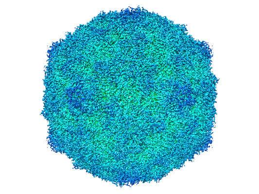 emd_11166.png emd_11166.png | 287.1 KB | ||
| Filedesc metadata | emd-11166.cif.gz | 5.9 KB | ||
| Archive directory |  http://ftp.pdbj.org/pub/emdb/structures/EMD-11166ftp://ftp.pdbj.org/pub/emdb/structures/EMD-11166 http://ftp.pdbj.org/pub/emdb/structures/EMD-11166ftp://ftp.pdbj.org/pub/emdb/structures/EMD-11166 | HTTPS FTP |
-Related structure data
| Related structure data |  6zclMC  6zckC 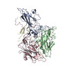 6zmsC M: atomic model generated by this map C: citing same article ( |
|---|---|
| Similar structure data |


-Links
| EMDB pages | EMDB (EBI/PDBe) / EMDataResource |
|---|---|
| Related items in Molecule of the Month |


-Map
| File | Download / File: emd_11166.map.gz / Format: CCP4 / Size: 282.6 MB / Type: IMAGE STORED AS FLOATING POINT NUMBER (4 BYTES) | ||||||||||||||||||||||||||||||||||||||||||||||||||||||||||||
|---|---|---|---|---|---|---|---|---|---|---|---|---|---|---|---|---|---|---|---|---|---|---|---|---|---|---|---|---|---|---|---|---|---|---|---|---|---|---|---|---|---|---|---|---|---|---|---|---|---|---|---|---|---|---|---|---|---|---|---|---|---|
| Annotation | Coxsackie B3 virion in complex with compound 17 | ||||||||||||||||||||||||||||||||||||||||||||||||||||||||||||
| Projections & slices | Image control
Images are generated by Spider. | ||||||||||||||||||||||||||||||||||||||||||||||||||||||||||||
| Voxel size | X=Y=Z: 1.06 Å | ||||||||||||||||||||||||||||||||||||||||||||||||||||||||||||
| Density |
| ||||||||||||||||||||||||||||||||||||||||||||||||||||||||||||
| Symmetry | Space group: 1 | ||||||||||||||||||||||||||||||||||||||||||||||||||||||||||||
| Details | EMDB XML:
CCP4 map header:
| ||||||||||||||||||||||||||||||||||||||||||||||||||||||||||||
 Z (Sec.)
 Y (Row.)
 X (Col.)


-Supplemental data
- Sample components
-Entire : Coxsackievirus B3 (strain Nancy)
| Entire | Name: Coxsackievirus B3 (strain Nancy) |
|---|---|
| Components |
|
-Supramolecule #1: Coxsackievirus B3 (strain Nancy)
| Supramolecule | Name: Coxsackievirus B3 (strain Nancy) / type: virus / ID: 1 / Parent: 0 / Macromolecule list: #1-#4 Details: Virus was grown in Vero A cells and purified in CsCl gradient NCBI-ID: 103903 / Sci species name: Coxsackievirus B3 (strain Nancy) / Sci species strain: Nancy / Virus type: VIRION / Virus isolate: STRAIN / Virus enveloped: No / Virus empty: No |
|---|---|
| Host (natural) | Organism:  human (human) human (human) |
| Virus shell | Shell ID: 1 / Name: icasaheadron / Diameter: 300.0 Å / T number (triangulation number): 3 |
-Macromolecule #1: capsid protein VP1
| Macromolecule | Name: capsid protein VP1 / type: protein_or_peptide / ID: 1 / Details: capsid protein VP1 / Number of copies: 1 / Enantiomer: LEVO / EC number: picornain 2A |
|---|---|
| Source (natural) | Organism: Coxsackievirus B3 (strain Nancy) |
| Molecular weight | Theoretical: 30.26893 KDa |
| Sequence | String: RVADTVGTGP TNSEAIPALT AAETGHTSQV VPGDTMQTRH VKNYHSRSES TIENFLCRSA CVYFTEYKNS GAKRYAEWVL TPRQAAQLR RKLEFFTYVR FDLELTFVIT STQQPSTTQN QDAQILTHQI MYVPPGGPVP DKVDSYVWQT STNPSVFWTE G NAPPRMSI ...String: RVADTVGTGP TNSEAIPALT AAETGHTSQV VPGDTMQTRH VKNYHSRSES TIENFLCRSA CVYFTEYKNS GAKRYAEWVL TPRQAAQLR RKLEFFTYVR FDLELTFVIT STQQPSTTQN QDAQILTHQI MYVPPGGPVP DKVDSYVWQT STNPSVFWTE G NAPPRMSI PFLSIGNAYS NFYDGWSEFS RNGVYGINTL NNMGTLYARH VNAGSTGPIK STIRIYFKPK HVKAWIPRPP RL CQYEKAK NVNFQPSGVT TTRQSITTMT NT UniProtKB: Genome polyprotein |
-Macromolecule #2: capsid protein VP2
| Macromolecule | Name: capsid protein VP2 / type: protein_or_peptide / ID: 2 / Details: capsid protein VP2 / Number of copies: 1 / Enantiomer: LEVO / EC number: picornain 2A |
|---|---|
| Source (natural) | Organism: Coxsackievirus B3 (strain Nancy) |
| Molecular weight | Theoretical: 27.604205 KDa |
| Sequence | String: SDRARSITLG NSTITTQECA NVVVGYGVWP DYLKDSEATA EDQPTQPDVA TCRFYTLDSV QWQKTSPGWW WKLPDALSNL GLFGQNMQY HYLGRTGYTV HVQCNASKFH QGCLLVVCVP EAEMGCATLD NTPSSAELLG GDTAKEFADK PVASGSNKLV Q RVVYNAGM ...String: SDRARSITLG NSTITTQECA NVVVGYGVWP DYLKDSEATA EDQPTQPDVA TCRFYTLDSV QWQKTSPGWW WKLPDALSNL GLFGQNMQY HYLGRTGYTV HVQCNASKFH QGCLLVVCVP EAEMGCATLD NTPSSAELLG GDTAKEFADK PVASGSNKLV Q RVVYNAGM GVGVGNLTIF PHQWINLRTN NSATIVMPYT NSVPMDNMFR HNNVTLMVIP FVPLDYCPGS TTYVPITVTI AP MCAEYNG LRLAG UniProtKB: Genome polyprotein |
-Macromolecule #3: capsid protein VP3
| Macromolecule | Name: capsid protein VP3 / type: protein_or_peptide / ID: 3 / Details: capsid protein VP3 / Number of copies: 1 / Enantiomer: LEVO / EC number: picornain 2A |
|---|---|
| Source (natural) | Organism: Coxsackievirus B3 (strain Nancy) |
| Molecular weight | Theoretical: 26.067596 KDa |
| Sequence | String: GLPTMNTPGS CQFLTSDDFQ SPSAMPQYDV TPEMRIPGEV KNLMEIAEVD SVVPVQNVGE KVNSMEAYQI PVRSNEGSGT QVFGFPLQP GYSSVFSRTL LGEILNYYTH WSGSIKLTFM FCGSAMATGK FLLAYSPPGA GAPTKRVDAM LGTHVIWDVG L QSSCVLCI ...String: GLPTMNTPGS CQFLTSDDFQ SPSAMPQYDV TPEMRIPGEV KNLMEIAEVD SVVPVQNVGE KVNSMEAYQI PVRSNEGSGT QVFGFPLQP GYSSVFSRTL LGEILNYYTH WSGSIKLTFM FCGSAMATGK FLLAYSPPGA GAPTKRVDAM LGTHVIWDVG L QSSCVLCI PWISQTHYRF VASDEYTAGG FITCWYQTNI VVPADAQSSC YIMCFVSACN DFSVRLLKDT PFISQQNFF UniProtKB: Genome polyprotein |
-Macromolecule #4: capsid protein VP4
| Macromolecule | Name: capsid protein VP4 / type: protein_or_peptide / ID: 4 / Details: myristoylated peptide, capsid protein VP4 / Number of copies: 1 / Enantiomer: LEVO / EC number: picornain 2A |
|---|---|
| Source (natural) | Organism: Coxsackievirus B3 (strain Nancy) |
| Molecular weight | Theoretical: 7.449181 KDa |
| Sequence | String: GAQVSTQKTG AHETRLNASG NSIIHYTNIN YYKDAASNSA NRQDFTQDPG KFTEPVKDIM IKSLPALN UniProtKB: Genome polyprotein |
-Macromolecule #5: 4-[[4-[1,3-bis(oxidanylidene)isoindol-2-yl]phenyl]sulfonylamino]b...
| Macromolecule | Name: 4-[[4-[1,3-bis(oxidanylidene)isoindol-2-yl]phenyl]sulfonylamino]benzoic acid type: ligand / ID: 5 / Number of copies: 1 / Formula: FHK |
|---|---|
| Molecular weight | Theoretical: 422.411 Da |
| Chemical component information | 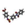 ChemComp-FHK: |
-Macromolecule #6: MYRISTIC ACID
| Macromolecule | Name: MYRISTIC ACID / type: ligand / ID: 6 / Number of copies: 1 / Formula: MYR |
|---|---|
| Molecular weight | Theoretical: 228.371 Da |
| Chemical component information |  ChemComp-MYR: |
-Experimental details
-Structure determination
| Method | cryo EM |
|---|---|
 Processing Processing | single particle reconstruction |
| Aggregation state | particle |
-Sample preparation
| Buffer | pH: 7.5 |
|---|---|
| Vitrification | Cryogen name: ETHANE / Instrument: HOMEMADE PLUNGER |
| Details | Purified virus was mixed with compound 17 and incubated at room temperature for 30 min before plunging |
- Electron microscopy
| Microscope | FEI TITAN KRIOS |
|---|---|
| Image recording | Film or detector model: GATAN K2 SUMMIT (4k x 4k) / Detector mode: COUNTING / Average electron dose: 47.0 e/Å2 |
| Electron beam | Acceleration voltage: 300 kV / Electron source:  FIELD EMISSION GUN FIELD EMISSION GUN |
| Electron optics | Illumination mode: FLOOD BEAM / Imaging mode: BRIGHT FIELD |
| Experimental equipment |  Model: Titan Krios / Image courtesy: FEI Company |
-Image processing
| Startup model | Type of model: INSILICO MODEL |
|---|---|
| Final reconstruction | Resolution.type: BY AUTHOR / Resolution: 2.8 Å / Resolution method: FSC 0.143 CUT-OFF / Number images used: 18626 |
| Initial angle assignment | Type: MAXIMUM LIKELIHOOD |
| Final angle assignment | Type: MAXIMUM LIKELIHOOD |
