 Movie
Movie Controller

[English] 日本語
 Yorodumi
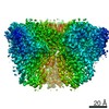
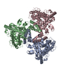
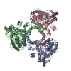
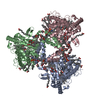
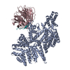
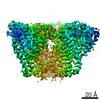
- PDB-6wr4: Structure of human ATG9A, the only transmembrane protein of the c... -
+ Open data

- Basic information
| Entry | Database: PDB / ID: 6wr4 | ||||||
|---|---|---|---|---|---|---|---|
| Title | Structure of human ATG9A, the only transmembrane protein of the core autophagy machinery | ||||||
 Components Components | Autophagy-related protein 9A | ||||||
 Keywords Keywords | MEMBRANE PROTEIN / TG9A / autophagosome / autophagy / cryoEM / molecular dynamics / transmembrane protein / membranecurvature / cellular compartments / membrane morphology / lipids | ||||||
| Function / homology |  Function and homology information Function and homology informationprogrammed necrotic cell death / phospholipid scramblase activity / protein localization to phagophore assembly site / phagophore assembly site membrane / piecemeal microautophagy of the nucleus / bone morphogenesis / phagophore assembly site / reticulophagy / Macroautophagy / autophagosome assembly ...programmed necrotic cell death / phospholipid scramblase activity / protein localization to phagophore assembly site / phagophore assembly site membrane / piecemeal microautophagy of the nucleus / bone morphogenesis / phagophore assembly site / reticulophagy / Macroautophagy / autophagosome assembly / mitophagy / autophagosome / synaptic membrane / PINK1-PRKN Mediated Mitophagy / trans-Golgi network / recycling endosome / mitochondrial membrane / recycling endosome membrane / late endosome / late endosome membrane / endosome / Golgi membrane / endoplasmic reticulum membrane / Golgi apparatus / mitochondrion / membrane Similarity search - Function | ||||||
| Biological species |  Homo sapiens (human) Homo sapiens (human) | ||||||
| Method | ELECTRON MICROSCOPY / single particle reconstruction / cryo EM / Resolution: 2.9 Å | ||||||
 Authors Authors | Guardia, C.M. / Tan, X. / Lian, T. / Rana, M.S. / Zhou, W. / Christenson, E.T. / Lowry, A.J. / Faraldo-Gomez, J.D. / Bonifacino, J.S. / Jiang, J. / Banerjee, A. | ||||||
 Citation Citation | Journal: Cell Rep / Year: 2020 Title: Structure of Human ATG9A, the Only Transmembrane Protein of the Core Autophagy Machinery. Authors: Carlos M Guardia / Xiao-Feng Tan / Tengfei Lian / Mitra S Rana / Wenchang Zhou / Eric T Christenson / Augustus J Lowry / José D Faraldo-Gómez / Juan S Bonifacino / Jiansen Jiang / Anirban Banerjee /  Abstract: Autophagy is a catabolic process involving capture of cytoplasmic materials into double-membraned autophagosomes that subsequently fuse with lysosomes for degradation of the materials by lysosomal ...Autophagy is a catabolic process involving capture of cytoplasmic materials into double-membraned autophagosomes that subsequently fuse with lysosomes for degradation of the materials by lysosomal hydrolases. One of the least understood components of the autophagy machinery is the transmembrane protein ATG9. Here, we report a cryoelectron microscopy structure of the human ATG9A isoform at 2.9-Å resolution. The structure reveals a fold with a homotrimeric domain-swapped architecture, multiple membrane spans, and a network of branched cavities, consistent with ATG9A being a membrane transporter. Mutational analyses support a role for the cavities in the function of ATG9A. In addition, structure-guided molecular simulations predict that ATG9A causes membrane bending, explaining the localization of this protein to small vesicles and highly curved edges of growing autophagosomes. | ||||||
| History |
|
- Structure visualization
| Movie |
Movie viewer |
|---|---|
| Structure viewer | Molecule: MolmilJmol/JSmol |
- Downloads & links
-Download
| PDBx/mmCIF format | 6wr4.cif.gz | 281.2 KB | Display | PDBx/mmCIF format |
|---|---|---|---|---|
| PDB format | pdb6wr4.ent.gz | 210.4 KB | Display | PDB format |
| PDBx/mmJSON format | 6wr4.json.gz | Tree view | PDBx/mmJSON format | |
| Others |  Other downloads Other downloads |
-Validation report
| Arichive directory | https://data.pdbj.org/pub/pdb/validation_reports/wr/6wr4ftp://data.pdbj.org/pub/pdb/validation_reports/wr/6wr4 | HTTPS FTP |
|---|
-Related structure data
| Related structure data | 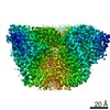 21876MC 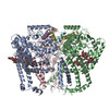 6wqzC M: map data used to model this data C: citing same article ( |
|---|---|
| Similar structure data |


-Links
 PDBj
PDBj


- Assembly
| Deposited unit | 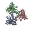
|
|---|---|
| 1 |
|
-Components
| #1: Protein | Mass: 94551.031 Da / Num. of mol.: 3 Source method: isolated from a genetically manipulated source Source: (gene. exp.) Homo sapiens (human) / Gene: ATG9A, APG9L1 / Production host: Homo sapiens (human) / References: UniProt: Q7Z3C6#2: Chemical | ChemComp-LMN /   Mass: 1005.188 Da / Num. of mol.: 6 / Source method: obtained synthetically / Formula: C47H88O22 / Feature type: SUBJECT OF INVESTIGATION / Comment: detergent*YM Mass: 1005.188 Da / Num. of mol.: 6 / Source method: obtained synthetically / Formula: C47H88O22 / Feature type: SUBJECT OF INVESTIGATION / Comment: detergent*YMHas ligand of interest | Y | |
|---|
-Experimental details
-Experiment
| Experiment | Method: ELECTRON MICROSCOPY |
|---|---|
| EM experiment | Aggregation state: PARTICLE / 3D reconstruction method: single particle reconstruction |
- Sample preparation
| Component | Name: Autophagy Related 9A with LMNG / Type: COMPLEX / Entity ID: #1 / Source: RECOMBINANT |
|---|---|
| Source (natural) | Organism: Homo sapiens (human) |
| Source (recombinant) | Organism: Homo sapiens (human) |
| Buffer solution | pH: 8 |
| Specimen | Embedding applied: NO / Shadowing applied: NO / Staining applied: NO / Vitrification applied: YES |
| Specimen support | Details: unspecified |
| Vitrification | Cryogen name: ETHANE |
- Electron microscopy imaging
| Experimental equipment |  Model: Titan Krios / Image courtesy: FEI Company |
|---|---|
| Microscopy | Model: FEI TITAN KRIOS |
| Electron gun | Electron source:  FIELD EMISSION GUN / Accelerating voltage: 300 kV / Illumination mode: FLOOD BEAM FIELD EMISSION GUN / Accelerating voltage: 300 kV / Illumination mode: FLOOD BEAM |
| Electron lens | Mode: BRIGHT FIELD / C2 aperture diameter: 70 µm |
| Image recording | Electron dose: 57 e/Å2 / Film or detector model: GATAN K2 SUMMIT (4k x 4k) |
- Processing
| Software |
| ||||||||||||
|---|---|---|---|---|---|---|---|---|---|---|---|---|---|
| CTF correction | Type: NONE | ||||||||||||
| 3D reconstruction | Resolution: 2.9 Å / Resolution method: FSC 0.143 CUT-OFF / Num. of particles: 646291 / Symmetry type: POINT | ||||||||||||
| Refinement | Cross valid method: THROUGHOUT | ||||||||||||
| Displacement parameters | Biso max: 0 Å2 / Biso mean: 0 Å2 / Biso min: 0 Å2 |
