 Movie
Movie Controller


+ Open data

- Basic information
| Entry | Database: PDB / ID: 6v4j | |||||||||||||||||||||
|---|---|---|---|---|---|---|---|---|---|---|---|---|---|---|---|---|---|---|---|---|---|---|
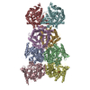
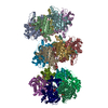
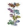
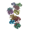
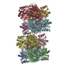
| Title | Structure of TrkH-TrkA in complex with ATP | |||||||||||||||||||||
 Components Components |
| |||||||||||||||||||||
 Keywords Keywords | TRANSPORT PROTEIN / Ion channel | |||||||||||||||||||||
| Function / homology |  Function and homology information Function and homology informationpotassium:chloride symporter activity / potassium ion transmembrane transporter activity / potassium ion binding / potassium channel activity / potassium ion transmembrane transport / nucleotide binding / protein homodimerization activity / metal ion binding / identical protein binding / plasma membrane Similarity search - Function | |||||||||||||||||||||
| Biological species | Vibrio parahaemolyticus serotype O3:K6 | |||||||||||||||||||||
| Method | ELECTRON MICROSCOPY / single particle reconstruction / cryo EM / Resolution: 2.97 Å | |||||||||||||||||||||
 Authors Authors | Zhou, M. / Zhang, H. | |||||||||||||||||||||
| Funding support |  United States, 2items United States, 2items
| |||||||||||||||||||||
 Citation Citation | Journal: Nat Commun / Year: 2020 Title: TrkA undergoes a tetramer-to-dimer conversion to open TrkH which enables changes in membrane potential. Authors: Hanzhi Zhang / Yaping Pan / Liya Hu / M Ashley Hudson / Katrina S Hofstetter / Zhichun Xu / Mingqiang Rong / Zhao Wang / B V Venkataram Prasad / Steve W Lockless / Wah Chiu / Ming Zhou / Abstract: TrkH is a bacterial ion channel implicated in K uptake and pH regulation. TrkH assembles with its regulatory protein, TrkA, which closes the channel when bound to ADP and opens it when bound to ATP. ...TrkH is a bacterial ion channel implicated in K uptake and pH regulation. TrkH assembles with its regulatory protein, TrkA, which closes the channel when bound to ADP and opens it when bound to ATP. However, it is unknown how nucleotides control the gating of TrkH through TrkA. Here we report the structures of the TrkH-TrkA complex in the presence of ADP or ATP. TrkA forms a tetrameric ring when bound to ADP and constrains TrkH to a closed conformation. The TrkA ring splits into two TrkA dimers in the presence of ATP and releases the constraints on TrkH, resulting in an open channel conformation. Functional studies show that both the tetramer-to-dimer conversion of TrkA and the loss of constraints on TrkH are required for channel gating. In addition, deletion of TrkA in Escherichia coli depolarizes the cell, suggesting that the TrkH-TrkA complex couples changes in intracellular nucleotides to membrane potential. | |||||||||||||||||||||
| History |
|
- Structure visualization
| Movie |
Movie viewer |
|---|---|
| Structure viewer | Molecule: MolmilJmol/JSmol |
- Downloads & links
-Download
| PDBx/mmCIF format | 6v4j.cif.gz | 584 KB | Display | PDBx/mmCIF format |
|---|---|---|---|---|
| PDB format | pdb6v4j.ent.gz | 477 KB | Display | PDB format |
| PDBx/mmJSON format | 6v4j.json.gz | Tree view | PDBx/mmJSON format | |
| Others |  Other downloads Other downloads |
-Validation report
| Arichive directory | https://data.pdbj.org/pub/pdb/validation_reports/v4/6v4jftp://data.pdbj.org/pub/pdb/validation_reports/v4/6v4j | HTTPS FTP |
|---|
-Related structure data
| Related structure data |  21041MC  6v4kC  6v4lC M: map data used to model this data C: citing same article ( |
|---|---|
| Similar structure data |
-Links
 PDBj
PDBj
- Assembly
| Deposited unit | 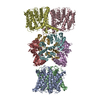
|
|---|---|
| 1 |
|
-Components
| #1: Protein | Mass: 53104.375 Da / Num. of mol.: 4 Source method: isolated from a genetically manipulated source Source: (gene. exp.)  Vibrio parahaemolyticus serotype O3:K6 (strain RIMD 2210633) (bacteria) Vibrio parahaemolyticus serotype O3:K6 (strain RIMD 2210633) (bacteria)Strain: RIMD 2210633 / Gene: trkH, VP0032 / Production host: #2: Protein | Mass: 50193.086 Da / Num. of mol.: 4 Source method: isolated from a genetically manipulated source Source: (gene. exp.) Vibrio parahaemolyticus serotype O3:K6 (strain RIMD 2210633) (bacteria)Strain: RIMD 2210633 / Gene: VP3045 / Production host: Has protein modification | Y | |
|---|
-Experimental details
-Experiment
| Experiment | Method: ELECTRON MICROSCOPY |
|---|---|
| EM experiment | Aggregation state: PARTICLE / 3D reconstruction method: single particle reconstruction |
- Sample preparation
| Component | Name: TrkH-TrkA / Type: COMPLEX / Entity ID: all / Source: RECOMBINANT |
|---|---|
| Molecular weight | Value: 0.4 MDa / Experimental value: NO |
| Source (natural) | Organism: Vibrio parahaemolyticus RIMD 2210633 (bacteria) |
| Source (recombinant) | Organism: |
| Buffer solution | pH: 7.5 |
| Specimen | Embedding applied: NO / Shadowing applied: NO / Staining applied: NO / Vitrification applied: YES |
| Specimen support | Details: unspecified |
| Vitrification | Cryogen name: ETHANE |
- Electron microscopy imaging
| Microscopy | Model: JEOL 3200FSC |
|---|---|
| Electron gun | Electron source:  FIELD EMISSION GUN / Accelerating voltage: 300 kV / Illumination mode: FLOOD BEAM FIELD EMISSION GUN / Accelerating voltage: 300 kV / Illumination mode: FLOOD BEAM |
| Electron lens | Mode: BRIGHT FIELD |
| Image recording | Electron dose: 80 e/Å2 / Film or detector model: GATAN K2 SUMMIT (4k x 4k) |
- Processing
| Software | Name: PHENIX / Version: 1.11.1_2575: / Classification: refinement | ||||||||||||||||||||||||
|---|---|---|---|---|---|---|---|---|---|---|---|---|---|---|---|---|---|---|---|---|---|---|---|---|---|
| EM software |
| ||||||||||||||||||||||||
| CTF correction | Type: PHASE FLIPPING AND AMPLITUDE CORRECTION | ||||||||||||||||||||||||
| 3D reconstruction | Resolution: 2.97 Å / Resolution method: FSC 0.143 CUT-OFF / Num. of particles: 72317 / Symmetry type: POINT | ||||||||||||||||||||||||
| Refine LS restraints |
|
