 Movie
Movie Controller


+ Open data

- Basic information
| Entry | Database: EMDB / ID: EMD-21041 | |||||||||
|---|---|---|---|---|---|---|---|---|---|---|
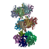
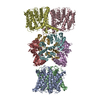
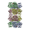
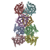
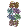
| Title | Structure of TrkH-TrkA in complex with ATP | |||||||||
 Map data Map data | Structure of TrkH-TrkA in complex with ATP | |||||||||
 Sample Sample |
| |||||||||
 Keywords Keywords | Ion channel / TRANSPORT PROTEIN | |||||||||
| Function / homology |  Function and homology information Function and homology informationpotassium:chloride symporter activity / potassium ion transmembrane transporter activity / potassium ion binding / potassium channel activity / potassium ion transmembrane transport / nucleotide binding / protein homodimerization activity / metal ion binding / identical protein binding / plasma membrane Similarity search - Function | |||||||||
| Biological species |  Vibrio parahaemolyticus RIMD 2210633 (bacteria) / Vibrio parahaemolyticus serotype O3:K6 (strain RIMD 2210633) (bacteria) Vibrio parahaemolyticus RIMD 2210633 (bacteria) / Vibrio parahaemolyticus serotype O3:K6 (strain RIMD 2210633) (bacteria) | |||||||||
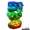
| Method | single particle reconstruction / cryo EM / Resolution: 2.97 Å | |||||||||
 Authors Authors | Zhou M / Zhang H | |||||||||
| Funding support |  United States, 2 items United States, 2 items
| |||||||||
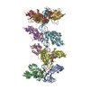
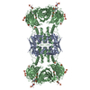
 Citation Citation | Journal: Nat Commun / Year: 2020 Title: TrkA undergoes a tetramer-to-dimer conversion to open TrkH which enables changes in membrane potential. Authors: Hanzhi Zhang / Yaping Pan / Liya Hu / M Ashley Hudson / Katrina S Hofstetter / Zhichun Xu / Mingqiang Rong / Zhao Wang / B V Venkataram Prasad / Steve W Lockless / Wah Chiu / Ming Zhou / Abstract: TrkH is a bacterial ion channel implicated in K uptake and pH regulation. TrkH assembles with its regulatory protein, TrkA, which closes the channel when bound to ADP and opens it when bound to ATP. ...TrkH is a bacterial ion channel implicated in K uptake and pH regulation. TrkH assembles with its regulatory protein, TrkA, which closes the channel when bound to ADP and opens it when bound to ATP. However, it is unknown how nucleotides control the gating of TrkH through TrkA. Here we report the structures of the TrkH-TrkA complex in the presence of ADP or ATP. TrkA forms a tetrameric ring when bound to ADP and constrains TrkH to a closed conformation. The TrkA ring splits into two TrkA dimers in the presence of ATP and releases the constraints on TrkH, resulting in an open channel conformation. Functional studies show that both the tetramer-to-dimer conversion of TrkA and the loss of constraints on TrkH are required for channel gating. In addition, deletion of TrkA in Escherichia coli depolarizes the cell, suggesting that the TrkH-TrkA complex couples changes in intracellular nucleotides to membrane potential. | |||||||||
| History |
|
- Structure visualization
| Movie |
Movie viewer |
|---|---|
| Structure viewer | EM map: SurfViewMolmilJmol/JSmol |
| Supplemental images |

- Downloads & links
-EMDB archive
| Map data | emd_21041.map.gz | 38 MB | EMDB map data format | |
|---|---|---|---|---|
| Header (meta data) | emd-21041-v30.xmlemd-21041.xml | 13.6 KB 13.6 KB | Display Display | EMDB header |
| Images |  emd_21041.png emd_21041.png | 118.9 KB | ||
| Filedesc metadata | emd-21041.cif.gz | 6.2 KB | ||
| Archive directory |  http://ftp.pdbj.org/pub/emdb/structures/EMD-21041ftp://ftp.pdbj.org/pub/emdb/structures/EMD-21041 http://ftp.pdbj.org/pub/emdb/structures/EMD-21041ftp://ftp.pdbj.org/pub/emdb/structures/EMD-21041 | HTTPS FTP |
-Related structure data
| Related structure data |  6v4jMC  6v4kC  6v4lC M: atomic model generated by this map C: citing same article ( |
|---|---|
| Similar structure data |
-Links
| EMDB pages | EMDB (EBI/PDBe) / EMDataResource |
|---|---|
| Related items in Molecule of the Month |

-Map
| File | Download / File: emd_21041.map.gz / Format: CCP4 / Size: 40.6 MB / Type: IMAGE STORED AS FLOATING POINT NUMBER (4 BYTES) | ||||||||||||||||||||||||||||||||||||||||||||||||||||||||||||||||||||
|---|---|---|---|---|---|---|---|---|---|---|---|---|---|---|---|---|---|---|---|---|---|---|---|---|---|---|---|---|---|---|---|---|---|---|---|---|---|---|---|---|---|---|---|---|---|---|---|---|---|---|---|---|---|---|---|---|---|---|---|---|---|---|---|---|---|---|---|---|---|
| Annotation | Structure of TrkH-TrkA in complex with ATP | ||||||||||||||||||||||||||||||||||||||||||||||||||||||||||||||||||||
| Projections & slices | Image control
Images are generated by Spider. | ||||||||||||||||||||||||||||||||||||||||||||||||||||||||||||||||||||
| Voxel size | X=Y=Z: 1.242 Å | ||||||||||||||||||||||||||||||||||||||||||||||||||||||||||||||||||||
| Density |
| ||||||||||||||||||||||||||||||||||||||||||||||||||||||||||||||||||||
| Symmetry | Space group: 1 | ||||||||||||||||||||||||||||||||||||||||||||||||||||||||||||||||||||
| Details | EMDB XML:
CCP4 map header:
| ||||||||||||||||||||||||||||||||||||||||||||||||||||||||||||||||||||
 Z (Sec.)
 Y (Row.)
 X (Col.)


-Supplemental data
- Sample components
-Entire : TrkH-TrkA
| Entire | Name: TrkH-TrkA |
|---|---|
| Components |
|
-Supramolecule #1: TrkH-TrkA
| Supramolecule | Name: TrkH-TrkA / type: complex / ID: 1 / Parent: 0 / Macromolecule list: all |
|---|---|
| Source (natural) | Organism: Vibrio parahaemolyticus RIMD 2210633 (bacteria) |
| Molecular weight | Theoretical: 400 KDa |
-Macromolecule #1: Trk system potassium uptake protein TrkH
| Macromolecule | Name: Trk system potassium uptake protein TrkH / type: protein_or_peptide / ID: 1 / Number of copies: 4 / Enantiomer: LEVO |
|---|---|
| Source (natural) | Organism: Vibrio parahaemolyticus serotype O3:K6 (strain RIMD 2210633) (bacteria) Strain: RIMD 2210633 |
| Molecular weight | Theoretical: 53.104375 KDa |
| Recombinant expression | Organism: |
| Sequence | String: MQFRSIIRIV GLLLALFSVT MLAPALVALL YRDGAGVPFV TTFFVLLFCG AMCWFPNRRH KHELKSRDGF LIVVLFWTVL GSAGSLPFL IADNPNISVT DAFFESFSAL TTTGATVIVG LDELPKAILF YRQFLQWFGG MGIIVLAVAI LPVLGIGGMQ L YRAEIPGP ...String: MQFRSIIRIV GLLLALFSVT MLAPALVALL YRDGAGVPFV TTFFVLLFCG AMCWFPNRRH KHELKSRDGF LIVVLFWTVL GSAGSLPFL IADNPNISVT DAFFESFSAL TTTGATVIVG LDELPKAILF YRQFLQWFGG MGIIVLAVAI LPVLGIGGMQ L YRAEIPGP VKDTKMTPRI AETAKALWYI YLSLTIACAV AFWLAGMTPF DAISHSFSTI AIGGFSTHDA SMGYFDSYAI NL ITVVFLL ISACNFTLHF AAFASGGVHP KYYWKDPEFR AFIFIQVLLF LVCFLLLLKH HSYTSPYDAF DQALFQTVSI STT AGFTTT GFADWPLFLP VLLLFSSFIG GCAGSTGGGM KVIRILLLTL QGARELKRLV HPRAVYTIKV GGSALPQRVV DAVW GFFSA YALVFVVCML GLIATGMDEL SAFSAVAATL NNLGPGLGEV ALHFGDVNDK AKWVLIVSML FGRLEIFTLL ILLTP TFWR S UniProtKB: Trk system potassium uptake protein TrkH |
-Macromolecule #2: Potassium uptake protein TrkA
| Macromolecule | Name: Potassium uptake protein TrkA / type: protein_or_peptide / ID: 2 / Number of copies: 4 / Enantiomer: LEVO |
|---|---|
| Source (natural) | Organism: Vibrio parahaemolyticus serotype O3:K6 (strain RIMD 2210633) (bacteria) Strain: RIMD 2210633 |
| Molecular weight | Theoretical: 50.193086 KDa |
| Recombinant expression | Organism: |
| Sequence | String: MKIIILGAGQ VGGTLAENLV GENNDITIVD NNADRLRELQ DKYDLRVVNG HASHPDVLHE AGAQDADMLV AVTNTDETNM AACQVAFTL FNTPNRVARI RSPEYLAEKE ALFKSGAIPV DHLIAPEELV TSYIERLIQY PGALQVVSFA EQKVSLVAVK A YYGGPLVG ...String: MKIIILGAGQ VGGTLAENLV GENNDITIVD NNADRLRELQ DKYDLRVVNG HASHPDVLHE AGAQDADMLV AVTNTDETNM AACQVAFTL FNTPNRVARI RSPEYLAEKE ALFKSGAIPV DHLIAPEELV TSYIERLIQY PGALQVVSFA EQKVSLVAVK A YYGGPLVG NALSALREHM PHIDTRVAAI FRQGRPIRPQ GTTIIEADDE VFFVAASNHI RSVMSELQRL EKPYRRIMIV GG GNIGASL AKRLEQTYSV KLIERDYQRA EKLSEQLENT IVFCGDAADQ ELLTEENIDQ VDVFIALTNE DETNIMSAML AKR MGAKKV MVLIQRGAYV DLVQGGVIDV AISPQQATIS ALLTHVRRAD IVNVSSLRRG AAEAIEAVAH GDETTSKVVG RAIG DIKLP PGTTIGAVVR GEEVLIAHDR TVIEQDDHVV MFLVDKKYVP DVEALFQPSP FFL UniProtKB: Trk system potassium uptake protein TrkA |
-Experimental details
-Structure determination
| Method | cryo EM |
|---|---|
 Processing Processing | single particle reconstruction |
| Aggregation state | particle |
-Sample preparation
| Buffer | pH: 7.5 |
|---|---|
| Grid | Details: unspecified |
| Vitrification | Cryogen name: ETHANE |
- Electron microscopy
| Microscope | JEOL 3200FSC |
|---|---|
| Image recording | Film or detector model: GATAN K2 SUMMIT (4k x 4k) / Average electron dose: 80.0 e/Å2 |
| Electron beam | Acceleration voltage: 300 kV / Electron source:  FIELD EMISSION GUN FIELD EMISSION GUN |
| Electron optics | Illumination mode: FLOOD BEAM / Imaging mode: BRIGHT FIELD |
