 Movie
Movie Controller


+ Open data

- Basic information
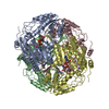
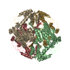
| Entry | Database: PDB / ID: 1bi9 | |||||||||
|---|---|---|---|---|---|---|---|---|---|---|
| Title | RETINAL DEHYDROGENASE TYPE TWO WITH NAD BOUND | |||||||||
 Components Components | RETINAL DEHYDROGENASE TYPE II | |||||||||
 Keywords Keywords | ALDEHYDE DEHYDROGENASE / RETINOID | |||||||||
| Function / homology |  Function and homology information Function and homology informationRA biosynthesis pathway / determination of bilateral symmetry / 9-cis-retinoic acid biosynthetic process / 3-chloroallyl aldehyde dehydrogenase activity / retinoic acid biosynthetic process / regulation of endothelial cell proliferation / embryonic camera-type eye development / retinal dehydrogenase / ureter maturation / midgut development ...RA biosynthesis pathway / determination of bilateral symmetry / 9-cis-retinoic acid biosynthetic process / 3-chloroallyl aldehyde dehydrogenase activity / retinoic acid biosynthetic process / regulation of endothelial cell proliferation / embryonic camera-type eye development / retinal dehydrogenase / ureter maturation / midgut development / proximal/distal pattern formation / morphogenesis of embryonic epithelium / pituitary gland development / aldehyde metabolic process / retinal metabolic process / hindbrain development / neural crest cell development / embryonic forelimb morphogenesis / embryonic digestive tract development / aldehyde dehydrogenase (NAD+) activity / anterior/posterior pattern specification / camera-type eye development / retinal binding / retinoic acid metabolic process / pancreas development / embryonic limb morphogenesis / retinal dehydrogenase (NAD+) activity / neural tube development / response to vitamin A / retinol metabolic process / cardiac muscle tissue development / forebrain development / face development / blood vessel development / response to retinoic acid / retinoic acid receptor signaling pathway / heart morphogenesis / cellular response to retinoic acid / lung development / response to cytokine / liver development / kidney development / neuron differentiation / response to estradiol / protein homotetramerization / positive regulation of apoptotic process / negative regulation of cell population proliferation / positive regulation of cell population proliferation / positive regulation of gene expression / perinuclear region of cytoplasm / cytoplasm Similarity search - Function | |||||||||
| Biological species |  | |||||||||
| Method |  X-RAY DIFFRACTION / MOLECULAR REPLACEMENT / Resolution: 2.7 Å X-RAY DIFFRACTION / MOLECULAR REPLACEMENT / Resolution: 2.7 Å | |||||||||
 Authors Authors | Newcomer, M.E. / Lamb, A.L. | |||||||||
 Citation Citation | Journal: Biochemistry / Year: 1999 Title: The structure of retinal dehydrogenase type II at 2.7 A resolution: implications for retinal specificity. Authors: Lamb, A.L. / Newcomer, M.E. #1: Journal: Acta Crystallogr.,Sect.D / Year: 1998Title: Purification, Crystallization and Preliminary X-Ray Diffraction Studies of Retinal Dehydrogenase Type II Authors: Lamb, A.L. / Wang, X. / Napoli, J.L. / Newcomer, M.E. #2: Journal: Mech.Dev. / Year: 1997Title: Restricted Expression and Retinoic Acid-Induced Downregulation of the Retinaldehyde Dehydrogenase Type 2 (Raldh-2) Gene During Mouse Development Authors: Niederreither, K. / Mccaffery, P. / Drager, U.C. / Chambon, P. / Dolle, P. #3: Journal: J.Biol.Chem. / Year: 1996Title: Cloning of a Cdna Encoding an Aldehyde Dehydrogenase and its Expression in Escherichia Coli. Recognition of Retinal as Substrate Authors: Wang, X. / Penzes, P. / Napoli, J.L. | |||||||||
| History |
|
- Structure visualization
| Structure viewer | Molecule: MolmilJmol/JSmol |
|---|
- Downloads & links
-Download
| PDBx/mmCIF format | 1bi9.cif.gz | 342.4 KB | Display | PDBx/mmCIF format |
|---|---|---|---|---|
| PDB format | pdb1bi9.ent.gz | 284.3 KB | Display | PDB format |
| PDBx/mmJSON format | 1bi9.json.gz | Tree view | PDBx/mmJSON format | |
| Others |  Other downloads Other downloads |
-Validation report
| Arichive directory | https://data.pdbj.org/pub/pdb/validation_reports/bi/1bi9ftp://data.pdbj.org/pub/pdb/validation_reports/bi/1bi9 | HTTPS FTP |
|---|
-Related structure data
| Related structure data | 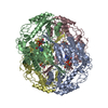 1a4zS S: Starting model for refinement |
|---|---|
| Similar structure data |


-Links
 PDBj
PDBj

- Assembly
| Deposited unit | 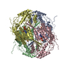
| ||||||||||||||||
|---|---|---|---|---|---|---|---|---|---|---|---|---|---|---|---|---|---|
| 1 |
| ||||||||||||||||
| 2 | 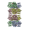
| ||||||||||||||||
| Unit cell |
| ||||||||||||||||
| Components on special symmetry positions |
| ||||||||||||||||
| Noncrystallographic symmetry (NCS) | NCS oper:
|
-Components
| #1: Protein | Mass: 54796.504 Da / Num. of mol.: 4 Source method: isolated from a genetically manipulated source Source: (gene. exp.)  References: UniProt: Q63639, Oxidoreductases; Acting on the aldehyde or oxo group of donors; With NAD+ or NADP+ as acceptor #2: Chemical | ChemComp-NAD /   Mass: 663.425 Da / Num. of mol.: 4 / Source method: obtained synthetically / Formula: C21H27N7O14P2 / Comment: NAD*YM Mass: 663.425 Da / Num. of mol.: 4 / Source method: obtained synthetically / Formula: C21H27N7O14P2 / Comment: NAD*YM#3: Chemical |   Mass: 35.453 Da / Num. of mol.: 2 / Source method: obtained synthetically / Formula: Cl Mass: 35.453 Da / Num. of mol.: 2 / Source method: obtained synthetically / Formula: Cl#4: Water | ChemComp-HOH / |  Mass: 18.015 Da / Num. of mol.: 224 / Source method: isolated from a natural source / Formula: H2O Mass: 18.015 Da / Num. of mol.: 224 / Source method: isolated from a natural source / Formula: H2OHas protein modification | N | |
|---|
-Experimental details
-Experiment
| Experiment | Method: X-RAY DIFFRACTION / Number of used crystals: 1 |
|---|
- Sample preparation
| Crystal | Density Matthews: 3.09 Å3/Da / Density % sol: 60 % | ||||||||||||||||||||||||||||||||||||||||||||||||||||||||||||||||||||||||
|---|---|---|---|---|---|---|---|---|---|---|---|---|---|---|---|---|---|---|---|---|---|---|---|---|---|---|---|---|---|---|---|---|---|---|---|---|---|---|---|---|---|---|---|---|---|---|---|---|---|---|---|---|---|---|---|---|---|---|---|---|---|---|---|---|---|---|---|---|---|---|---|---|---|
| Crystal grow | pH: 7.1 / Details: pH 7.1 | ||||||||||||||||||||||||||||||||||||||||||||||||||||||||||||||||||||||||
| Crystal grow | *PLUS Temperature: 25 ℃ / pH: 8.5 / Method: vapor diffusion, sitting drop | ||||||||||||||||||||||||||||||||||||||||||||||||||||||||||||||||||||||||
| Components of the solutions | *PLUS
|
-Data collection
| Diffraction | Mean temperature: 123 K |
|---|---|
| Diffraction source | Source: ROTATING ANODE / Type: RIGAKU RUH2R / Wavelength: 1.5418 |
| Detector | Type: RIGAKU RAXIS II / Detector: IMAGE PLATE / Date: Sep 1, 1997 / Details: MIRRORS |
| Radiation | Monochromator: NI FILTER / Monochromatic (M) / Laue (L): M / Scattering type: x-ray |
| Radiation wavelength | Wavelength: 1.5418 Å / Relative weight: 1 |
| Reflection | Resolution: 2.5→30 Å / Num. obs: 75572 / % possible obs: 99.7 % / Observed criterion σ(I): 2 / Redundancy: 4.8 % / Biso Wilson estimate: 53.2 Å2 / Rmerge(I) obs: 0.075 / Net I/σ(I): 22 |
| Reflection shell | Resolution: 2.7→2.8 Å / Redundancy: 4.8 % / Rmerge(I) obs: 0.412 / Mean I/σ(I) obs: 3 / % possible all: 99.9 |
| Reflection | *PLUS Num. measured all: 864923 |
| Reflection shell | *PLUS % possible obs: 99.9 % / Mean I/σ(I) obs: 3.6 |
- Processing
| Software |
| ||||||||||||||||||||||||||||||||||||||||||||||||||||||||||||
|---|---|---|---|---|---|---|---|---|---|---|---|---|---|---|---|---|---|---|---|---|---|---|---|---|---|---|---|---|---|---|---|---|---|---|---|---|---|---|---|---|---|---|---|---|---|---|---|---|---|---|---|---|---|---|---|---|---|---|---|---|---|
| Refinement | Method to determine structure: MOLECULAR REPLACEMENT Starting model: 1A4Z Resolution: 2.7→30 Å / Rfactor Rfree error: 0.003 / Data cutoff high absF: 283265.58 / Data cutoff low absF: 0 / Isotropic thermal model: RESTRAINED / Cross valid method: THROUGHOUT / σ(F): 0
| ||||||||||||||||||||||||||||||||||||||||||||||||||||||||||||
| Solvent computation | Solvent model: FLAT MODEL / Bsol: 42.49 Å2 / ksol: 0.326 e/Å3 | ||||||||||||||||||||||||||||||||||||||||||||||||||||||||||||
| Displacement parameters | Biso mean: 48.1 Å2
| ||||||||||||||||||||||||||||||||||||||||||||||||||||||||||||
| Refine analyze |
| ||||||||||||||||||||||||||||||||||||||||||||||||||||||||||||
| Refinement step | Cycle: LAST / Resolution: 2.7→30 Å /
| ||||||||||||||||||||||||||||||||||||||||||||||||||||||||||||
| Refine LS restraints |
| ||||||||||||||||||||||||||||||||||||||||||||||||||||||||||||
| LS refinement shell | Resolution: 2.7→2.87 Å / Rfactor Rfree error: 0.012 / Total num. of bins used: 6
| ||||||||||||||||||||||||||||||||||||||||||||||||||||||||||||
| Xplor file |
| ||||||||||||||||||||||||||||||||||||||||||||||||||||||||||||
| Software | *PLUS Name: CNS / Version: 0.4 / Classification: refinement | ||||||||||||||||||||||||||||||||||||||||||||||||||||||||||||
| Refine LS restraints | *PLUS
|
