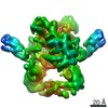
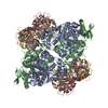
登録情報 データベース : PDB / ID : 3gutタイトル Crystal structure of a higher-order complex of p50:RelA bound to the HIV-1 LTR HIV-LTR Core Forward Strand HIV-LTR Core Reverse Strand Nuclear factor NF-kappa-B p105 subunit Transcription factor p65 キーワード / / / / / / / / / / / / / / / / / / / / 機能・相同性 分子機能 ドメイン・相同性 構成要素
/ / / / / / / / / / / / / / / / / / / / / / / / / / / / / / / / / / / / / / / / / / / / / / / / / / / / / / / / / / / / / / / / / / / / / / / / / / / / / / / / / / / / / / / / / / / / / / / / / / / / / / / / / / / / / / / / / / / / / / / / / / / / / / / / / / / / / / / / / / / / / / / / / / / / / / / / 生物種 Homo sapiens (ヒト)手法 / / / 解像度 : 3.59 Å データ登録者 Stroud, J.C. / Oltman, A.J. / Han, A. / Bates, D.L. / Chen, L. ジャーナル : J.Mol.Biol. / 年 : 2009タイトル : Structural basis of HIV-1 activation by NF-kappaB--a higher-order complex of p50:RelA bound to the HIV-1 LTR.著者 : Stroud, J.C. / Oltman, A. / Han, A. / Bates, D.L. / Chen, L. 履歴 登録 2009年3月30日 登録サイト / 処理サイト 改定 1.0 2009年9月8日 Provider / タイプ 改定 1.1 2011年7月13日 Group 改定 1.2 2017年11月1日 Group / カテゴリ 改定 1.3 2023年9月6日 Group / Database references / Refinement descriptionカテゴリ chem_comp_atom / chem_comp_bond ... chem_comp_atom / chem_comp_bond / database_2 / pdbx_initial_refinement_model / struct_ref_seq_dif Item / _database_2.pdbx_database_accession / _struct_ref_seq_dif.details
すべて表示 表示を減らす
 ムービー
ムービー コントローラー


 データを開く
 基本情報
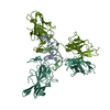
基本情報 要素
要素 キーワード
キーワード 機能・相同性情報
 Homo sapiens (ヒト)
 Human immunodeficiency virus (ヒト免疫不全ウイルス)
 X線回折 /
  データ登録者
 引用
引用 構造の表示
構造の表示 ダウンロードとリンク
 その他のダウンロード
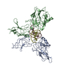


 PDBj
PDBj


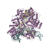
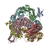
 集合体
集合体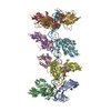


 試料調製
試料調製 / ビームライン: 8.2.2 / 波長: 1.0688 Å
 解析
解析