photosystem I reaction center / photosystem I / photosynthetic electron transport in photosystem I / photosystem I / plasma membrane-derived thylakoid membrane / chlorophyll binding / photosynthesis / 4 iron, 4 sulfur cluster binding / electron transfer activity / oxidoreductase activity ...photosystem I reaction center / photosystem I / photosynthetic electron transport in photosystem I / photosystem I / plasma membrane-derived thylakoid membrane / chlorophyll binding / photosynthesis / 4 iron, 4 sulfur cluster binding / electron transfer activity / oxidoreductase activity / magnesium ion binding / metal ion binding / plasma membrane Similarity search - Function
Photosystem 1 Reaction Centre Subunit Xi; Chain: L; / Photosystem I PsaL, reaction centre subunit XI / Photosystem I reaction center subunit PsaK / Photosystem I reaction centre subunit PsaK / Photosystem I reaction centre subunit PsaK superfamily / Photosystem I psaG and psaK proteins signature. / Photosystem I PsaM, reaction centre superfamily / Photosystem I PsaM, reaction centre / Photosystem I protein M (PsaM) / Photosystem I reaction center subunit V/PsaK ...Photosystem 1 Reaction Centre Subunit Xi; Chain: L; / Photosystem I PsaL, reaction centre subunit XI / Photosystem I reaction center subunit PsaK / Photosystem I reaction centre subunit PsaK / Photosystem I reaction centre subunit PsaK superfamily / Photosystem I psaG and psaK proteins signature. / Photosystem I PsaM, reaction centre superfamily / Photosystem I PsaM, reaction centre / Photosystem I protein M (PsaM) / Photosystem I reaction center subunit V/PsaK / Photosystem I psaG / psaK / Photosystem I PsaL, reaction centre subunit XI / Photosystem I, reaction centre subunit XI / Photosystem I PsaL, reaction centre subunit XI superfamily / Photosystem I reaction centre subunit XI / Photosystem I reaction centre subunit VIII / Photosystem I reaction centre subunit VIII / Photosystem I reaction centre subunit VIII superfamily / Photosystem I PsaF, reaction centre subunit III / Photosystem I PsaF, reaction centre subunit III superfamily / Photosystem I reaction centre subunit III / Photosystem I PsaD / Photosystem I, reaction centre subunit PsaD superfamily / PsaD / Photosystem I PsaE, reaction centre subunit IV / Photosystem I reaction centre subunit IV / PsaE / Photosystem I PsaJ, reaction centre subunit IX superfamily / Photosystem I PsaJ, reaction centre subunit IX / Photosystem I reaction centre subunit IX / PsaJ / Photosystem I PsaA / Photosystem I protein PsaC / Photosystem I PsaB / Photosystem I PsaA/PsaB, conserved site / Photosystem I psaA and psaB proteins signature. / : / Photosystem I PsaA/PsaB / Photosystem I PsaA/PsaB superfamily / Photosystem I psaA/psaB protein / Electron transport accessory-like domain superfamily / 4Fe-4S dicluster domain / 4Fe-4S ferredoxin, iron-sulphur binding, conserved site / 4Fe-4S ferredoxin-type iron-sulfur binding region signature. / 4Fe-4S ferredoxin-type iron-sulfur binding domain profile. / 4Fe-4S ferredoxin-type, iron-sulphur binding domain / Up-down Bundle / Mainly Alpha Similarity search - Domain/homology
BETA-CAROTENE / CHLOROPHYLL A ISOMER / CHLOROPHYLL A / 1,2-DIPALMITOYL-PHOSPHATIDYL-GLYCEROLE / 1,2-DISTEAROYL-MONOGALACTOSYL-DIGLYCERIDE / PHYLLOQUINONE / IRON/SULFUR CLUSTER / Photosystem I reaction center subunit IV / Photosystem I reaction center subunit II / Photosystem I P700 chlorophyll a apoprotein A1 ...BETA-CAROTENE / CHLOROPHYLL A ISOMER / CHLOROPHYLL A / 1,2-DIPALMITOYL-PHOSPHATIDYL-GLYCEROLE / 1,2-DISTEAROYL-MONOGALACTOSYL-DIGLYCERIDE / PHYLLOQUINONE / IRON/SULFUR CLUSTER / Photosystem I reaction center subunit IV / Photosystem I reaction center subunit II / Photosystem I P700 chlorophyll a apoprotein A1 / Photosystem I P700 chlorophyll a apoprotein A2 / Photosystem I reaction center subunit III / Photosystem I iron-sulfur center / Photosystem I reaction center subunit XI / Photosystem I reaction center subunit XII / Photosystem I reaction center subunit PsaK 2 / Photosystem I reaction center subunit IX / Photosystem I reaction center subunit VIII Similarity search - Component
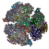
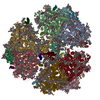
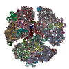
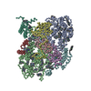
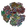
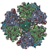
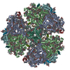
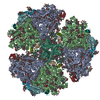
Journal: Nat Commun / Year: 2020 Title: The structure of a red-shifted photosystem I reveals a red site in the core antenna. Authors: Hila Toporik / Anton Khmelnitskiy / Zachary Dobson / Reece Riddle / Dewight Williams / Su Lin / Ryszard Jankowiak / Yuval Mazor / Abstract: Photosystem I coordinates more than 90 chlorophylls in its core antenna while achieving near perfect quantum efficiency. Low energy chlorophylls (also known as red chlorophylls) residing in the ...Photosystem I coordinates more than 90 chlorophylls in its core antenna while achieving near perfect quantum efficiency. Low energy chlorophylls (also known as red chlorophylls) residing in the antenna are important for energy transfer dynamics and yield, however, their precise location remained elusive. Here, we construct a chimeric Photosystem I complex in Synechocystis PCC 6803 that shows enhanced absorption in the red spectral region. We combine Cryo-EM and spectroscopy to determine the structurefunction relationship in this red-shifted Photosystem I complex. Determining the structure of this complex reveals the precise architecture of the low energy site as well as large scale structural heterogeneity which is probably universal to all trimeric Photosystem I complexes. Identifying the structural elements that constitute red sites can expand the absorption spectrum of oxygenic photosynthetic and potentially modulate light harvesting efficiency.
A: Photosystem I P700 chlorophyll a apoprotein A1 B: Photosystem I P700 chlorophyll a apoprotein A2 C: Photosystem I iron-sulfur center D: Photosystem I reaction center subunit II E: Photosystem I reaction center subunit IV F: Photosystem I reaction center subunit III I: Photosystem I reaction center subunit VIII J: Photosystem I reaction center subunit IX L: Photosystem I reaction center subunit XI M: Photosystem I reaction center subunit XII K: Photosystem I reaction center subunit PsaK 2 a: Photosystem I P700 chlorophyll a apoprotein A1 1: Photosystem I P700 chlorophyll a apoprotein A1 c: Photosystem I iron-sulfur center 3: Photosystem I iron-sulfur center b: Photosystem I P700 chlorophyll a apoprotein A2 2: Photosystem I P700 chlorophyll a apoprotein A2 d: Photosystem I reaction center subunit II 4: Photosystem I reaction center subunit II e: Photosystem I reaction center subunit IV 5: Photosystem I reaction center subunit IV f: Photosystem I reaction center subunit III 6: Photosystem I reaction center subunit III i: Photosystem I reaction center subunit VIII h: Photosystem I reaction center subunit VIII j: Photosystem I reaction center subunit IX 7: Photosystem I reaction center subunit IX k: Photosystem I reaction center subunit PsaK 2 8: Photosystem I reaction center subunit PsaK 2 l: Photosystem I reaction center subunit XI 0: Photosystem I reaction center subunit XI m: Photosystem I reaction center subunit XII 9: Photosystem I reaction center subunit XII hetero molecules
 Movie
Movie Controller


 Open data
 Basic information
 Components
 Keywords
 Function and homology information
 Authors
 Citation
 Structure visualization
 UCSF Chimera
 Downloads & links
 Other downloads
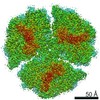
 PDBj
PDBj


 Assembly