Movie
Movie Controller

[English] 日本語
 Yorodumi
- PDB-6qm5: Cryo-EM structure of calcium-bound nhTMEM16 lipid scramblase in DDM -
+ Open data

- Basic information
| Entry | Database: PDB / ID: 6qm5 | |||||||||
|---|---|---|---|---|---|---|---|---|---|---|
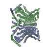
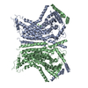
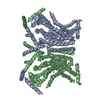
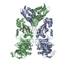
| Title | Cryo-EM structure of calcium-bound nhTMEM16 lipid scramblase in DDM | |||||||||
 Components Components | Predicted protein | |||||||||
 Keywords Keywords | MEMBRANE PROTEIN / lipid scrambles / TMEM16 | |||||||||
| Function / homology |  Function and homology information Function and homology informationcortical endoplasmic reticulum / chloride channel activity / membrane / metal ion binding / identical protein binding Similarity search - Function | |||||||||
| Biological species |  Nectria haematococca (fungus) Nectria haematococca (fungus) | |||||||||
| Method | ELECTRON MICROSCOPY / single particle reconstruction / cryo EM / Resolution: 3.6 Å | |||||||||
 Authors Authors | Kalienkova, V. / Clerico Mosina, V. / Bryner, L. / Oostergetel, G.T. / Dutzler, R. / Paulino, C. | |||||||||
| Funding support |  Switzerland, Switzerland,  Netherlands, 2items Netherlands, 2items
| |||||||||
 Citation Citation | Journal: Elife / Year: 2019 Title: Stepwise activation mechanism of the scramblase nhTMEM16 revealed by cryo-EM. Authors: Valeria Kalienkova / Vanessa Clerico Mosina / Laura Bryner / Gert T Oostergetel / Raimund Dutzler / Cristina Paulino / Abstract: Scramblases catalyze the movement of lipids between both leaflets of a bilayer. Whereas the X-ray structure of the protein nhTMEM16 has previously revealed the architecture of a Ca-dependent lipid ...Scramblases catalyze the movement of lipids between both leaflets of a bilayer. Whereas the X-ray structure of the protein nhTMEM16 has previously revealed the architecture of a Ca-dependent lipid scramblase, its regulation mechanism has remained elusive. Here, we have used cryo-electron microscopy and functional assays to address this question. Ca-bound and Ca-free conformations of nhTMEM16 in detergent and lipid nanodiscs illustrate the interactions with its environment and they reveal the conformational changes underlying its activation. In this process, Ca binding induces a stepwise transition of the catalytic subunit cavity, converting a closed cavity that is shielded from the membrane in the absence of ligand, into a polar furrow that becomes accessible to lipid headgroups in the Ca-bound state. Additionally, our structures demonstrate how nhTMEM16 distorts the membrane at both entrances of the subunit cavity, thereby decreasing the energy barrier for lipid movement. | |||||||||
| History |
|
- Structure visualization
| Movie |
Movie viewer |
|---|---|
| Structure viewer | Molecule: MolmilJmol/JSmol |
- Downloads & links
-Download
| PDBx/mmCIF format | 6qm5.cif.gz | 245 KB | Display | PDBx/mmCIF format |
|---|---|---|---|---|
| PDB format | pdb6qm5.ent.gz | 197.3 KB | Display | PDB format |
| PDBx/mmJSON format | 6qm5.json.gz | Tree view | PDBx/mmJSON format | |
| Others |  Other downloads Other downloads |
-Validation report
| Arichive directory | https://data.pdbj.org/pub/pdb/validation_reports/qm/6qm5ftp://data.pdbj.org/pub/pdb/validation_reports/qm/6qm5 | HTTPS FTP |
|---|
-Related structure data
| Related structure data | 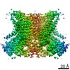 4588MC 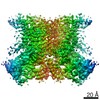 4587C 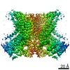 4589C 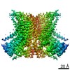 4592C 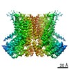 4593C 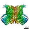 4594C 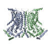 6qm4C  6qm6C 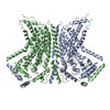 6qm9C  6qmaC 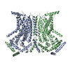 6qmbC M: map data used to model this data C: citing same article ( |
|---|---|
| Similar structure data |

-Links
 PDBj
PDBj- Assembly
| Deposited unit | 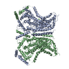
|
|---|---|
| 1 |
|
-Components
| #1: Protein | Mass: 83200.008 Da / Num. of mol.: 2 Source method: isolated from a genetically manipulated source Source: (gene. exp.) Nectria haematococca (fungus) / Gene: NECHADRAFT_66456 / Production host:  #2: Chemical | ChemComp-CA /   Mass: 40.078 Da / Num. of mol.: 4 / Source method: obtained synthetically / Formula: Ca / Feature type: SUBJECT OF INVESTIGATION Mass: 40.078 Da / Num. of mol.: 4 / Source method: obtained synthetically / Formula: Ca / Feature type: SUBJECT OF INVESTIGATION |
|---|
-Experimental details
-Experiment
| Experiment | Method: ELECTRON MICROSCOPY |
|---|---|
| EM experiment | Aggregation state: PARTICLE / 3D reconstruction method: single particle reconstruction |
- Sample preparation
| Component | Name: nhTMEM16 / Type: COMPLEX / Entity ID: #1 / Source: RECOMBINANT |
|---|---|
| Molecular weight | Value: 0.166 MDa / Experimental value: NO |
| Source (natural) | Organism: Nectria haematococca mpVI 77-13-4 (fungus) |
| Source (recombinant) | Organism: |
| Buffer solution | pH: 7.6 Details: 5 mM Hepes 7.6, 150 mM NaCl, 0.03% DDM, 2 mM EGTA. 2.3 mM CaCl2 were added 30 minutes before freezing. |
| Specimen | Conc.: 3.3 mg/ml / Embedding applied: NO / Shadowing applied: NO / Staining applied: NO / Vitrification applied: YES |
| Specimen support | Details: at 5 mA / Grid material: GOLD / Grid mesh size: 300 divisions/in. / Grid type: Quantifoil R1.2/1.3 |
| Vitrification | Instrument: FEI VITROBOT MARK IV / Cryogen name: ETHANE / Humidity: 100 % / Chamber temperature: 288 K |
- Electron microscopy imaging
| Experimental equipment |  Model: Talos Arctica / Image courtesy: FEI Company |
|---|---|
| Microscopy | Model: FEI TALOS ARCTICA |
| Electron gun | Electron source:  FIELD EMISSION GUN / Accelerating voltage: 200 kV / Illumination mode: FLOOD BEAM FIELD EMISSION GUN / Accelerating voltage: 200 kV / Illumination mode: FLOOD BEAM |
| Electron lens | Mode: BRIGHT FIELD / Nominal magnification: 49407 X / Calibrated magnification: 49407 X / Nominal defocus max: 3000 nm / Nominal defocus min: 300 nm / Calibrated defocus min: 300 nm / Calibrated defocus max: 3000 nm / Cs: 2.7 mm / C2 aperture diameter: 100 µm / Alignment procedure: COMA FREE |
| Specimen holder | Cryogen: NITROGEN / Specimen holder model: FEI TITAN KRIOS AUTOGRID HOLDER / Temperature (max): 105 K / Temperature (min): 90 K |
| Image recording | Average exposure time: 9 sec. / Electron dose: 52 e/Å2 / Detector mode: COUNTING / Film or detector model: GATAN K2 SUMMIT (4k x 4k) / Num. of grids imaged: 9 / Num. of real images: 2521 |
| EM imaging optics | Energyfilter name: GIF Bioquantum / Energyfilter slit width: 20 eV |
| Image scans | Width: 3838 / Height: 3710 / Movie frames/image: 60 / Used frames/image: 1-60 |
- Processing
| Software | Name: PHENIX / Version: 1.14_3260: / Classification: refinement | ||||||||||||||||||||||||||||||||||||
|---|---|---|---|---|---|---|---|---|---|---|---|---|---|---|---|---|---|---|---|---|---|---|---|---|---|---|---|---|---|---|---|---|---|---|---|---|---|
| EM software |
| ||||||||||||||||||||||||||||||||||||
| CTF correction | Type: PHASE FLIPPING AND AMPLITUDE CORRECTION | ||||||||||||||||||||||||||||||||||||
| Particle selection | Num. of particles selected: 251693 | ||||||||||||||||||||||||||||||||||||
| Symmetry | Point symmetry: C2 (2 fold cyclic) | ||||||||||||||||||||||||||||||||||||
| 3D reconstruction | Resolution: 3.6 Å / Resolution method: FSC 0.143 CUT-OFF / Num. of particles: 120086 / Algorithm: BACK PROJECTION / Symmetry type: POINT | ||||||||||||||||||||||||||||||||||||
| Atomic model building | Space: REAL | ||||||||||||||||||||||||||||||||||||
| Atomic model building | PDB-ID: 4WIS Accession code: 4WIS / Source name: PDB / Type: experimental model |