 Movie
Movie Controller

[English] 日本語
 Yorodumi
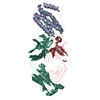
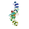
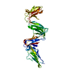
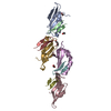
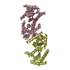
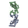
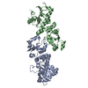
- PDB-6n3a: SegA-long, conformation of TDP-43 low complexity domain segment A long -
+ Open data

- Basic information
| Entry | Database: PDB / ID: 6n3a | ||||||
|---|---|---|---|---|---|---|---|
| Title | SegA-long, conformation of TDP-43 low complexity domain segment A long | ||||||
 Components Components |
| ||||||
 Keywords Keywords | dna binding protein / protein fibril / Amyloid / TDP43 / ALS / FTLD-TDP | ||||||
| Function / homology |  Function and homology information Function and homology informationnuclear inner membrane organization / interchromatin granule / perichromatin fibrils / 3'-UTR-mediated mRNA destabilization / 3'-UTR-mediated mRNA stabilization / negative regulation of protein phosphorylation / host-mediated suppression of viral transcription / pre-mRNA intronic binding / RNA splicing / response to endoplasmic reticulum stress ...nuclear inner membrane organization / interchromatin granule / perichromatin fibrils / 3'-UTR-mediated mRNA destabilization / 3'-UTR-mediated mRNA stabilization / negative regulation of protein phosphorylation / host-mediated suppression of viral transcription / pre-mRNA intronic binding / RNA splicing / response to endoplasmic reticulum stress / mRNA 3'-UTR binding / molecular condensate scaffold activity / regulation of protein stability / regulation of circadian rhythm / positive regulation of protein import into nucleus / mRNA processing / cytoplasmic stress granule / positive regulation of insulin secretion / rhythmic process / regulation of gene expression / double-stranded DNA binding / regulation of apoptotic process / amyloid fibril formation / regulation of cell cycle / nuclear speck / RNA polymerase II cis-regulatory region sequence-specific DNA binding / negative regulation of gene expression / lipid binding / chromatin / mitochondrion / DNA binding / RNA binding / nucleoplasm / identical protein binding / nucleus Similarity search - Function | ||||||
| Biological species |  Homo sapiens (human) Homo sapiens (human) | ||||||
| Method | ELECTRON MICROSCOPY / helical reconstruction / cryo EM / Resolution: 3.3 Å | ||||||
 Authors Authors | Cao, Q. / Boyer, D.R. / Sawaya, M.R. / Eisenberg, D.S. | ||||||
| Funding support |  United States, 1items United States, 1items
| ||||||
 Citation Citation | Journal: Nat Struct Mol Biol / Year: 2019 Title: Cryo-EM structures of four polymorphic TDP-43 amyloid cores. Authors: Qin Cao / David R Boyer / Michael R Sawaya / Peng Ge / David S Eisenberg / Abstract: The DNA and RNA processing protein TDP-43 undergoes both functional and pathogenic aggregation. Functional TDP-43 aggregates form reversible, transient species such as nuclear bodies, stress ...The DNA and RNA processing protein TDP-43 undergoes both functional and pathogenic aggregation. Functional TDP-43 aggregates form reversible, transient species such as nuclear bodies, stress granules, and myo-granules. Pathogenic, irreversible TDP-43 aggregates form in amyotrophic lateral sclerosis and other neurodegenerative conditions. Here we find the features of TDP-43 fibrils that confer both reversibility and irreversibility by determining structures of two segments reported to be the pathogenic cores of human TDP-43 aggregation: SegA (residues 311-360), which forms three polymorphs, all with dagger-shaped folds; and SegB A315E (residues 286-331 containing the amyotrophic lateral sclerosis hereditary mutation A315E), which forms R-shaped folds. Energetic analysis suggests that the dagger-shaped polymorphs represent irreversible fibril structures, whereas the SegB polymorph may participate in both reversible and irreversible fibrils. Our structures reveal the polymorphic nature of TDP-43 and suggest how the A315E mutation converts the R-shaped polymorph to an irreversible form that enhances pathology. | ||||||
| History |
|
- Structure visualization
| Movie |
Movie viewer |
|---|---|
| Structure viewer | Molecule: MolmilJmol/JSmol |
- Downloads & links
-Download
| PDBx/mmCIF format | 6n3a.cif.gz | 92.8 KB | Display | PDBx/mmCIF format |
|---|---|---|---|---|
| PDB format | pdb6n3a.ent.gz | 74.7 KB | Display | PDB format |
| PDBx/mmJSON format | 6n3a.json.gz | Tree view | PDBx/mmJSON format | |
| Others |  Other downloads Other downloads |
-Validation report
| Arichive directory | https://data.pdbj.org/pub/pdb/validation_reports/n3/6n3aftp://data.pdbj.org/pub/pdb/validation_reports/n3/6n3a | HTTPS FTP |
|---|
-Related structure data
| Related structure data |  9349MC  0334C  9339C  9350C  6n37C  6n3bC  6n3cC C: citing same article ( M: map data used to model this data |
|---|---|
| Similar structure data | |
| EM raw data | EMPIAR-10497 (Title: Cryo-EM structures of four polymorphic TDP-43 amyloid cores Data size: 4.5 TB Data #1: Unaligned dose fractionated frames of TDP-43 SegA amyloid fibrils [micrographs - multiframe] Data #2: Unaligned dose fractionated frames of TDP-43 SegB A315E amyloid fibrils [micrographs - multiframe]) |

-Links
 PDBj
PDBj
- Assembly
| Deposited unit | 
|
|---|---|
| 1 |
|
-Components
| #1: Protein/peptide | Mass: 5207.797 Da / Num. of mol.: 10 Source method: isolated from a genetically manipulated source Source: (gene. exp.) Homo sapiens (human) / Gene: TARDBP, TDP43 / Plasmid: pET28a / Production host:  #2: Protein/peptide | Mass: 1063.143 Da / Num. of mol.: 10 Source method: isolated from a genetically manipulated source Source: (gene. exp.) Homo sapiens (human) / Production host: |
|---|
-Experimental details
-Experiment
| Experiment | Method: ELECTRON MICROSCOPY |
|---|---|
| EM experiment | Aggregation state: FILAMENT / 3D reconstruction method: helical reconstruction |
- Sample preparation
| Component | Name: TDP43 fibril / Type: ORGANELLE OR CELLULAR COMPONENT / Entity ID: all / Source: RECOMBINANT |
|---|---|
| Source (natural) | Organism: Homo sapiens (human) |
| Source (recombinant) | Organism: |
| Buffer solution | pH: 7.4 |
| Specimen | Embedding applied: NO / Shadowing applied: NO / Staining applied: NO / Vitrification applied: YES |
| Vitrification | Cryogen name: ETHANE |
- Electron microscopy imaging
| Experimental equipment |  Model: Titan Krios / Image courtesy: FEI Company |
|---|---|
| Microscopy | Model: FEI TITAN KRIOS |
| Electron gun | Electron source:  FIELD EMISSION GUN / Accelerating voltage: 300 kV / Illumination mode: FLOOD BEAM FIELD EMISSION GUN / Accelerating voltage: 300 kV / Illumination mode: FLOOD BEAM |
| Electron lens | Mode: BRIGHT FIELD / Nominal defocus max: 2 nm / Nominal defocus min: 2 nm / Cs: 2.7 mm / C2 aperture diameter: 50 µm |
| Image recording | Average exposure time: 8 sec. / Electron dose: 48 e/Å2 / Detector mode: SUPER-RESOLUTION / Film or detector model: GATAN K2 SUMMIT (4k x 4k) |
- Processing
| EM software |
| ||||||||||||||||||||||||||||||||||||||||
|---|---|---|---|---|---|---|---|---|---|---|---|---|---|---|---|---|---|---|---|---|---|---|---|---|---|---|---|---|---|---|---|---|---|---|---|---|---|---|---|---|---|
| CTF correction | Type: PHASE FLIPPING AND AMPLITUDE CORRECTION | ||||||||||||||||||||||||||||||||||||||||
| Helical symmerty | Angular rotation/subunit: 179.34 ° / Axial rise/subunit: 4.84 Å / Axial symmetry: C2 | ||||||||||||||||||||||||||||||||||||||||
| 3D reconstruction | Resolution: 3.3 Å / Resolution method: FSC 0.143 CUT-OFF / Num. of particles: 167087 / Symmetry type: HELICAL | ||||||||||||||||||||||||||||||||||||||||
| Atomic model building | Protocol: AB INITIO MODEL |
