- PDB-5ebc: Crystal structure of EccB1 of Mycobacterium tuberculosis in space... -
+
Open data
ID or keywords:
Loading...
-
Basic information
Entry
Database: PDB / ID: 5ebc
Title
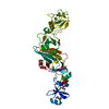
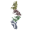
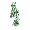
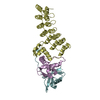
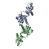
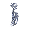
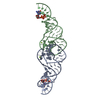
Crystal structure of EccB1 of Mycobacterium tuberculosis in spacegroup P21 (state III)
Components
ESX-1 secretion system protein eccB1
Keywords
PROTEIN TRANSPORT / ALPHA-BETA-ALPHA SANDWICH / BETA-SHEET / ATPASE / T7SS / ESX-1 SECRETION SYSTEM
Function / homology
Function and homology information
symbiont-mediated perturbation of host innate immune response / Hydrolases; Acting on acid anhydrides / peptidoglycan-based cell wall / hydrolase activity / extracellular region / ATP binding / plasma membrane Similarity search - Function
Type VII secretion system EccB, repeat 3 domain / Type VII secretion system EccB, repeat 1 domain / TTHA1013/TTHA0281-like / Type VII secretion system EccB / Type VII secretion system EccB, repeat 3 domain / Type VII secretion system EccB, repeat 1 domain / Type VII secretion system ESX-1, transport TM domain B / OB fold (Dihydrolipoamide Acetyltransferase, E2P) / Beta Barrel / 2-Layer Sandwich ...Type VII secretion system EccB, repeat 3 domain / Type VII secretion system EccB, repeat 1 domain / TTHA1013/TTHA0281-like / Type VII secretion system EccB / Type VII secretion system EccB, repeat 3 domain / Type VII secretion system EccB, repeat 1 domain / Type VII secretion system ESX-1, transport TM domain B / OB fold (Dihydrolipoamide Acetyltransferase, E2P) / Beta Barrel / 2-Layer Sandwich / Mainly Beta / Alpha Beta Similarity search - Domain/homology
In the structure databanks used in Yorodumi, some data are registered as the other names, "COVID-19 virus" and "2019-nCoV". Here are the details of the virus and the list of structure data.
Jan 31, 2019. EMDB accession codes are about to change! (news from PDBe EMDB page)
EMDB accession codes are about to change! (news from PDBe EMDB page)
The allocation of 4 digits for EMDB accession codes will soon come to an end. Whilst these codes will remain in use, new EMDB accession codes will include an additional digit and will expand incrementally as the available range of codes is exhausted. The current 4-digit format prefixed with “EMD-” (i.e. EMD-XXXX) will advance to a 5-digit format (i.e. EMD-XXXXX), and so on. It is currently estimated that the 4-digit codes will be depleted around Spring 2019, at which point the 5-digit format will come into force.
The EM Navigator/Yorodumi systems omit the EMD- prefix.
Related info.:Q: What is EMD? / ID/Accession-code notation in Yorodumi/EM Navigator
Yorodumi is a browser for structure data from EMDB, PDB, SASBDB, etc.
This page is also the successor to EM Navigator detail page, and also detail information page/front-end page for Omokage search.
The word "yorodu" (or yorozu) is an old Japanese word meaning "ten thousand". "mi" (miru) is to see.
Related info.:EMDB / PDB / SASBDB / Comparison of 3 databanks / Yorodumi Search / Aug 31, 2016. New EM Navigator & Yorodumi / Yorodumi Papers / Jmol/JSmol / Function and homology information / Changes in new EM Navigator and Yorodumi
 Movie
Movie Controller

 Yorodumi
 Open data
 Basic information
 Components
 Keywords
 Function and homology information
 Mycobacterium tuberculosis (bacteria)
 X-RAY DIFFRACTION /
  Authors
 Citation
 Structure visualization
 Downloads & links
 Other downloads


 PDBj
PDBj Assembly
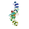

 Mass: 40.078 Da / Num. of mol.: 1 / Source method: obtained synthetically / Formula: Ca
 Sample preparation
 Processing