 Movie
Movie Controller


+ Open data

- Basic information
| Entry | Database: PDB / ID: 6mxf | ||||||||||||||||||
|---|---|---|---|---|---|---|---|---|---|---|---|---|---|---|---|---|---|---|---|
| Title | MicroED structure of thiostrepton at 1.9 A resolution | ||||||||||||||||||
 Components Components | Thiostrepton | ||||||||||||||||||
 Keywords Keywords | ANTIBIOTIC | ||||||||||||||||||
| Function / homology | Thiazolylpeptide-type bacteriocin precursor / Thiopeptide-type bacteriocin precursor / defense response to bacterium / extracellular region / THIOSTREPTON / Thiostrepton Function and homology information Function and homology information | ||||||||||||||||||
| Biological species |  Streptomyces azureus (bacteria) Streptomyces azureus (bacteria) | ||||||||||||||||||
| Method | ELECTRON CRYSTALLOGRAPHY / electron crystallography /  MOLECULAR REPLACEMENT / cryo EM / Resolution: 1.91 Å MOLECULAR REPLACEMENT / cryo EM / Resolution: 1.91 Å | ||||||||||||||||||
 Authors Authors | Jones, C.G. / Martynowycz, M.W. / Hattne, J. / Fulton, T. / Stoltz, B.M. / Rodriguez, J.A. / Nelson, H.M. / Gonen, T. | ||||||||||||||||||
| Funding support |  United States, 5items United States, 5items
| ||||||||||||||||||
 Citation Citation | Journal: ACS Cent Sci / Year: 2018 Title: The CryoEM Method MicroED as a Powerful Tool for Small Molecule Structure Determination. Authors: Christopher G Jones / Michael W Martynowycz / Johan Hattne / Tyler J Fulton / Brian M Stoltz / Jose A Rodriguez / Hosea M Nelson / Tamir Gonen / Abstract: In the many scientific endeavors that are driven by organic chemistry, unambiguous identification of small molecules is of paramount importance. Over the past 50 years, NMR and other powerful ...In the many scientific endeavors that are driven by organic chemistry, unambiguous identification of small molecules is of paramount importance. Over the past 50 years, NMR and other powerful spectroscopic techniques have been developed to address this challenge. While almost all of these techniques rely on inference of connectivity, the unambiguous determination of a small molecule's structure requires X-ray and/or neutron diffraction studies. In practice, however, X-ray crystallography is rarely applied in routine organic chemistry due to intrinsic limitations of both the analytes and the technique. Here we report the use of the electron cryo-microscopy (cryoEM) method microcrystal electron diffraction (MicroED) to provide routine and unambiguous structural determination of small organic molecules. From simple powders, with minimal sample preparation, we could collect high-quality MicroED data from nanocrystals (∼100 nm, ∼10 g) resulting in atomic resolution (<1 Å) crystal structures in minutes. | ||||||||||||||||||
| History |
|
- Structure visualization
| Movie |
Movie viewer |
|---|---|
| Structure viewer | Molecule: MolmilJmol/JSmol |
- Downloads & links
-Download
| PDBx/mmCIF format | 6mxf.cif.gz | 17.9 KB | Display | PDBx/mmCIF format |
|---|---|---|---|---|
| PDB format | pdb6mxf.ent.gz | 9.4 KB | Display | PDB format |
| PDBx/mmJSON format | 6mxf.json.gz | Tree view | PDBx/mmJSON format | |
| Others |  Other downloads Other downloads |
-Validation report
| Arichive directory | https://data.pdbj.org/pub/pdb/validation_reports/mx/6mxfftp://data.pdbj.org/pub/pdb/validation_reports/mx/6mxf | HTTPS FTP |
|---|
-Related structure data
| Related structure data |  9292MC  9282C  9283C  9284C 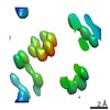 9285C 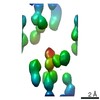 9286C 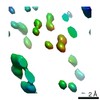 9287C 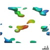 9288C 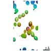 9289C 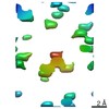 9290C 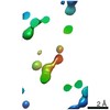 9291C 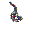 1e9wS |
|---|---|
| Similar structure data |


-Links
 PDBj
PDBj- Assembly
| Deposited unit | 
| ||||||||
|---|---|---|---|---|---|---|---|---|---|
| 1 |
| ||||||||
| Unit cell |
|
-Components
| #1: Protein/peptide | 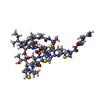  Type: Thiopeptide / Class: Antibiotic / Mass: 1805.985 Da / Num. of mol.: 1 / Source method: obtained synthetically Type: Thiopeptide / Class: Antibiotic / Mass: 1805.985 Da / Num. of mol.: 1 / Source method: obtained syntheticallyDetails: Thiostrepton is a hetrocyclic thiopeptide belonging to the thiocillin family, consisting of four thiazole, one thiozoline and one piperideine rings. A modified quinoline linked to main-chain ...Details: Thiostrepton is a hetrocyclic thiopeptide belonging to the thiocillin family, consisting of four thiazole, one thiozoline and one piperideine rings. A modified quinoline linked to main-chain residue 1 and side-chain of residue 12. Post translational maturation of thiazole and oxazole containing antibiotics involves the enzymic condensation of a Cys or Ser with the alpha-carbonyl of the preceding amino acid to form a thioether or ether bond, then dehydration to form a double bond with the alpha-amino nitrogen. Thiazoline or oxazoline ring are dehydrogenated to form thiazole or oxazole rings. the pyridinyl involves the cross-linking of a Ser and a Cys-Ser pair usually separated by 7 or 8 residues along the peptide chain. The Ser residues are dehydrated to didehydroalanines, then bonded between their beta carbons. The alpha carbonyl of the Cys condenses with alpha carbon of the first Ser to form a pyridinyl ring. The ring may be mutiply dehydrogenated to form a pyridine ring with loss of the amino nitrogen of the first Ser. The amidation of Ser-17 probably does not occur by the same mechanism, oxidative cleavage of glycine, as in eukaryotes. Source: (synth.) Streptomyces azureus (bacteria) / References: THIOSTREPTON, UniProt: P0C8P8*PLUS |
|---|---|
| #2: Water | ChemComp-HOH /  Mass: 18.015 Da / Num. of mol.: 2 / Source method: isolated from a natural source / Formula: H2O Mass: 18.015 Da / Num. of mol.: 2 / Source method: isolated from a natural source / Formula: H2O |
| Compound details | THIOSTREPTON IS A MEMBER OF A SULPHUR-RICH HETEROCYCLIC PEPTIDES CLASS. ALL SHARE A MACROCYLIC ...THIOSTREPT |
-Experimental details
-Experiment
| Experiment | Method: ELECTRON CRYSTALLOGRAPHY |
|---|---|
| EM experiment | Aggregation state: 3D ARRAY / 3D reconstruction method: electron crystallography |
- Sample preparation
| Component | Name: Thiostrepton / Type: ORGANELLE OR CELLULAR COMPONENT / Entity ID: #1 / Source: NATURAL |
|---|---|
| Molecular weight | Value: .001619538000 MDa / Experimental value: NO |
| Source (natural) | Organism: Streptomyces azureus (bacteria) |
| EM crystal formation | Details: Powder |
| Buffer solution | pH: 7 / Details: Powder |
| Specimen | Embedding applied: NO / Shadowing applied: NO / Staining applied: NO / Vitrification applied: YES / Details: Powder |
| Specimen support | Grid material: COPPER / Grid mesh size: 300 divisions/in. / Grid type: Quantifoil R2/2 |
| Vitrification | Cryogen name: NITROGEN / Humidity: 100 % / Chamber temperature: 298 K / Details: Hand-plunged |
-Data collection
| Experimental equipment |  Model: Talos Arctica / Image courtesy: FEI Company |
|---|---|
| Microscopy | Model: FEI TALOS ARCTICA |
| Electron gun | Electron source: FIELD EMISSION GUN / Accelerating voltage: 200 kV / Illumination mode: FLOOD BEAM |
| Electron lens | Mode: DIFFRACTION |
| Specimen holder | Cryogen: NITROGEN / Specimen holder model: FEI TITAN KRIOS AUTOGRID HOLDER |
| Image recording | Average exposure time: 2.21 sec. / Electron dose: 0.09 e/Å2 / Film or detector model: FEI CETA (4k x 4k) / Num. of diffraction images: 214 / Num. of grids imaged: 1 / Details: FEI CetaD |
| Image scans | Sampling size: 28 µm / Width: 2048 / Height: 2048 |
| EM diffraction | Camera length: 960 mm |
| EM diffraction shell | Resolution: 1.91→2.13 Å / Fourier space coverage: 40.3 % / Multiplicity: 4.9 / Num. of structure factors: 92 / Phase residual: 29.19 ° |
| EM diffraction stats | Fourier space coverage: 78.6 % / High resolution: 1.91 Å / Num. of intensities measured: 5578 / Num. of structure factors: 686 / Phase error: 26.93 ° / Phase residual: 26.93 ° / Phase error rejection criteria: 0 / Rmerge: 0.236 / Rsym: 0.236 |
| Diffraction source | Wavelength: 0.0251 Å |
| Detector | Date: Oct 3, 2018 |
| Radiation wavelength | Wavelength: 0.0251 Å / Relative weight: 1 |
| Reflection | Resolution: 1.9→18.99 Å / Num. obs: 686 / % possible obs: 78.6 % / Redundancy: 8.1 % / CC1/2: 0.985 / Rmerge(I) obs: 0.236 / Rpim(I) all: 0.084 / Rrim(I) all: 0.251 / Net I/σ(I): 5.1 |
| Reflection shell | Resolution: 1.9→2.13 Å / Redundancy: 4.9 % / Rmerge(I) obs: 0.32 / Mean I/σ(I) obs: 3.4 / Num. unique all: 93 / CC1/2: 0.813 / % possible all: 40.3 |
- Processing
| Software |
| ||||||||||||||||||||||||||||||||||||||||||||||||||||||||||||||||||||||||||||||||||||||||||||||||||||||||||
|---|---|---|---|---|---|---|---|---|---|---|---|---|---|---|---|---|---|---|---|---|---|---|---|---|---|---|---|---|---|---|---|---|---|---|---|---|---|---|---|---|---|---|---|---|---|---|---|---|---|---|---|---|---|---|---|---|---|---|---|---|---|---|---|---|---|---|---|---|---|---|---|---|---|---|---|---|---|---|---|---|---|---|---|---|---|---|---|---|---|---|---|---|---|---|---|---|---|---|---|---|---|---|---|---|---|---|---|
| EM software |
| ||||||||||||||||||||||||||||||||||||||||||||||||||||||||||||||||||||||||||||||||||||||||||||||||||||||||||
| Image processing | Details: FEI Ceta | ||||||||||||||||||||||||||||||||||||||||||||||||||||||||||||||||||||||||||||||||||||||||||||||||||||||||||
| EM 3D crystal entity | ∠α: 90 ° / ∠β: 90 ° / ∠γ: 90 ° / A: 26.219 Å / B: 26.219 Å / C: 27.534 Å / Space group name: P43212 / Space group num: 96 | ||||||||||||||||||||||||||||||||||||||||||||||||||||||||||||||||||||||||||||||||||||||||||||||||||||||||||
| CTF correction | Type: NONE | ||||||||||||||||||||||||||||||||||||||||||||||||||||||||||||||||||||||||||||||||||||||||||||||||||||||||||
| 3D reconstruction | Resolution: 1.91 Å / Resolution method: DIFFRACTION PATTERN/LAYERLINES / Symmetry type: 3D CRYSTAL | ||||||||||||||||||||||||||||||||||||||||||||||||||||||||||||||||||||||||||||||||||||||||||||||||||||||||||
| Atomic model building | B value: 2.6 / Protocol: OTHER / Space: RECIPROCAL | ||||||||||||||||||||||||||||||||||||||||||||||||||||||||||||||||||||||||||||||||||||||||||||||||||||||||||
| Atomic model building | PDB-ID: 1E9W Pdb chain-ID: A / Accession code: 1E9W / Pdb chain residue range: 0-18 / Source name: PDB / Type: experimental model | ||||||||||||||||||||||||||||||||||||||||||||||||||||||||||||||||||||||||||||||||||||||||||||||||||||||||||
| Refinement | Method to determine structure: MOLECULAR REPLACEMENT Starting model: 1e9w Resolution: 1.91→18.99 Å / Cor.coef. Fo:Fc: 0.931 / Cor.coef. Fo:Fc free: 0.94 / SU B: 3.282 / SU ML: 0.098 / Cross valid method: THROUGHOUT / ESU R: 0.352 / ESU R Free: 0.202 / Details: HYDROGENS HAVE BEEN ADDED IN THE RIDING POSITIONS
| ||||||||||||||||||||||||||||||||||||||||||||||||||||||||||||||||||||||||||||||||||||||||||||||||||||||||||
| Solvent computation | Ion probe radii: 0.8 Å / Shrinkage radii: 0.8 Å / VDW probe radii: 1.2 Å | ||||||||||||||||||||||||||||||||||||||||||||||||||||||||||||||||||||||||||||||||||||||||||||||||||||||||||
| Displacement parameters | Biso mean: 6.811 Å2
| ||||||||||||||||||||||||||||||||||||||||||||||||||||||||||||||||||||||||||||||||||||||||||||||||||||||||||
| Refinement step | Cycle: 1 / Total: 116 | ||||||||||||||||||||||||||||||||||||||||||||||||||||||||||||||||||||||||||||||||||||||||||||||||||||||||||
| Refine LS restraints |
|