 Movie
Movie Controller


+ Open data

- Basic information
| Entry | Database: EMDB / ID: EMD-9283 | ||||||||||||||||||
|---|---|---|---|---|---|---|---|---|---|---|---|---|---|---|---|---|---|---|---|
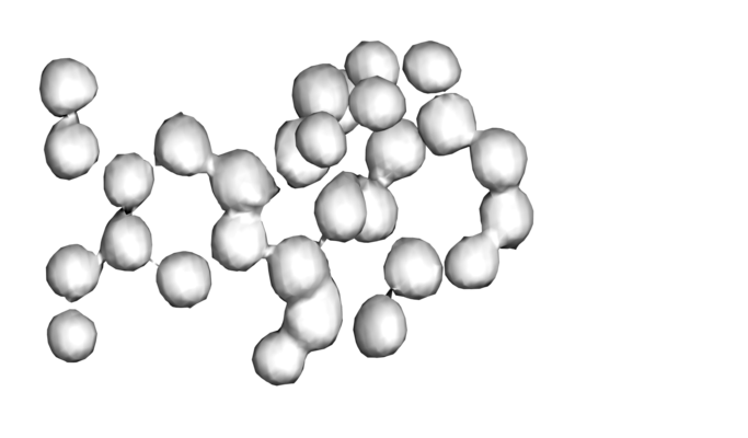
| Title | MicroED map of brucine at 0.9 A resolution | ||||||||||||||||||
 Map data Map data | 2Fo-Fc map of brucine | ||||||||||||||||||
 Sample Sample |
| ||||||||||||||||||
| Biological species | synthetic construct (others) | ||||||||||||||||||
| Method | electron crystallography / cryo EM | ||||||||||||||||||
 Authors Authors | Jones CG / Martynowycz MW / Hattne J / Fulton T / Stoltz BM / Rodriguez JA / Nelson HM / Gonen T | ||||||||||||||||||
| Funding support |  United States, 5 items United States, 5 items
| ||||||||||||||||||
 Citation Citation | Journal: ACS Cent Sci / Year: 2018 Title: The CryoEM Method MicroED as a Powerful Tool for Small Molecule Structure Determination. Authors: Christopher G Jones / Michael W Martynowycz / Johan Hattne / Tyler J Fulton / Brian M Stoltz / Jose A Rodriguez / Hosea M Nelson / Tamir Gonen / Abstract: In the many scientific endeavors that are driven by organic chemistry, unambiguous identification of small molecules is of paramount importance. Over the past 50 years, NMR and other powerful ...In the many scientific endeavors that are driven by organic chemistry, unambiguous identification of small molecules is of paramount importance. Over the past 50 years, NMR and other powerful spectroscopic techniques have been developed to address this challenge. While almost all of these techniques rely on inference of connectivity, the unambiguous determination of a small molecule's structure requires X-ray and/or neutron diffraction studies. In practice, however, X-ray crystallography is rarely applied in routine organic chemistry due to intrinsic limitations of both the analytes and the technique. Here we report the use of the electron cryo-microscopy (cryoEM) method microcrystal electron diffraction (MicroED) to provide routine and unambiguous structural determination of small organic molecules. From simple powders, with minimal sample preparation, we could collect high-quality MicroED data from nanocrystals (∼100 nm, ∼10 g) resulting in atomic resolution (<1 Å) crystal structures in minutes. | ||||||||||||||||||
| History |
|
- Structure visualization
| Movie |
 Movie viewer Movie viewer |
|---|---|
| Structure viewer | EM map: SurfViewMolmilJmol/JSmol |
| Supplemental images |
- Downloads & links
-EMDB archive
| Map data | emd_9283.map.gz | 196.4 KB | EMDB map data format | |
|---|---|---|---|---|
| Header (meta data) | emd-9283-v30.xmlemd-9283.xml | 14.1 KB 14.1 KB | Display Display | EMDB header |
| Images |  emd_9283.png emd_9283.png | 62.6 KB | ||
| Archive directory |  http://ftp.pdbj.org/pub/emdb/structures/EMD-9283ftp://ftp.pdbj.org/pub/emdb/structures/EMD-9283 http://ftp.pdbj.org/pub/emdb/structures/EMD-9283ftp://ftp.pdbj.org/pub/emdb/structures/EMD-9283 | HTTPS FTP |
-Related structure data
| Related structure data | 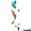 9282C 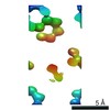 9284C 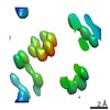 9285C 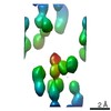 9286C 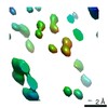 9287C 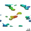 9288C 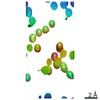 9289C 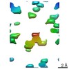 9290C 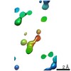 9291C  9292C  6mxfC C: citing same article ( |
|---|
-Links
| EMDB pages | EMDB (EBI/PDBe) / EMDataResource |
|---|
-Map
| File | Download / File: emd_9283.map.gz / Format: CCP4 / Size: 212.9 KB / Type: IMAGE STORED AS FLOATING POINT NUMBER (4 BYTES) | ||||||||||||||||||||||||||||||||||||||||||||||||||||||||||||||||||||
|---|---|---|---|---|---|---|---|---|---|---|---|---|---|---|---|---|---|---|---|---|---|---|---|---|---|---|---|---|---|---|---|---|---|---|---|---|---|---|---|---|---|---|---|---|---|---|---|---|---|---|---|---|---|---|---|---|---|---|---|---|---|---|---|---|---|---|---|---|---|
| Annotation | 2Fo-Fc map of brucine | ||||||||||||||||||||||||||||||||||||||||||||||||||||||||||||||||||||
| Projections & slices | Image control
Images are generated by Spider. generated in cubic-lattice coordinate | ||||||||||||||||||||||||||||||||||||||||||||||||||||||||||||||||||||
| Voxel size | X: 0.28407 Å / Y: 0.26929 Å / Z: 0.27792 Å | ||||||||||||||||||||||||||||||||||||||||||||||||||||||||||||||||||||
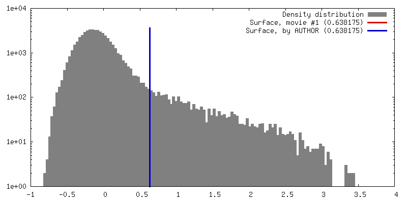
| Density |
| ||||||||||||||||||||||||||||||||||||||||||||||||||||||||||||||||||||
| Symmetry | Space group: 4 | ||||||||||||||||||||||||||||||||||||||||||||||||||||||||||||||||||||
| Details | EMDB XML:
CCP4 map header:
| ||||||||||||||||||||||||||||||||||||||||||||||||||||||||||||||||||||
 Z (Sec.)
 X (Row.)
 Y (Col.)


-Supplemental data
- Sample components
-Entire : Brucine
| Entire | Name: Brucine |
|---|---|
| Components |
|
-Supramolecule #1: Brucine
| Supramolecule | Name: Brucine / type: organelle_or_cellular_component / ID: 1 / Parent: 0 / Macromolecule list: #1 |
|---|---|
| Source (natural) | Organism: synthetic construct (others) |
| Molecular weight | Theoretical: 362.275 Da |
-Experimental details
-Structure determination
| Method | cryo EM |
|---|---|
 Processing Processing | electron crystallography |
| Aggregation state | 3D array |
-Sample preparation
| Concentration | 1 mg/mL |
|---|---|
| Buffer | pH: 7 / Details: Powder |
| Grid | Details: unspecified |
| Vitrification | Cryogen name: NITROGEN / Chamber humidity: 100 % / Chamber temperature: 298 K / Details: Hand-plunged. |
| Details | Powder |
| Crystal formation | Details: Powder |
- Electron microscopy
| Microscope | FEI TALOS ARCTICA |
|---|---|
| Image recording | Film or detector model: FEI CETA (4k x 4k) / Digitization - Dimensions - Width: 2048 pixel / Digitization - Dimensions - Height: 2048 pixel / Digitization - Sampling interval: 28.0 µm / Number grids imaged: 1 / Number diffraction images: 236 / Average exposure time: 3.05 sec. / Average electron dose: 0.09 e/Å2 / Details: FEI CetaD |
| Electron beam | Acceleration voltage: 200 kV / Electron source:  FIELD EMISSION GUN FIELD EMISSION GUN |
| Electron optics | Illumination mode: FLOOD BEAM / Imaging mode: DIFFRACTION / Camera length: 670 mm |
| Sample stage | Specimen holder model: FEI TITAN KRIOS AUTOGRID HOLDER / Cooling holder cryogen: NITROGEN |
| Experimental equipment |  Model: Talos Arctica / Image courtesy: FEI Company |
-Image processing
| Final reconstruction | Resolution method: DIFFRACTION PATTERN/LAYERLINES |
|---|---|
| Crystal parameters | Unit cell - A: 15.34 Å / Unit cell - B: 7.54 Å / Unit cell - C: 20.01 Å / Unit cell - γ: 90 ° / Unit cell - α: 90 ° / Unit cell - β: 112.49 ° / Space group: P 21 |
| Crystallography statistics | Number intensities measured: 12427 / Number structure factors: 5858 / Fourier space coverage: 95.3 / R sym: 0.182 / R merge: 0.242 / Overall phase error: 49.68 / Overall phase residual: 49.68 / Phase error rejection criteria: 0 / High resolution: 0.9 Å / Shell - Shell ID: 1 / Shell - High resolution: 0.9 Å / Shell - Low resolution: 0.923 Å / Shell - Number structure factors: 225 / Shell - Phase residual: 63.25 / Shell - Fourier space coverage: 97.4 / Shell - Multiplicity: 1.99 |
-Atomic model buiding 1
| Refinement | Space: RECIPROCAL / Protocol: AB INITIO MODEL |
|---|
