Movie
Movie Controller


+ Open data

- Basic information
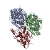
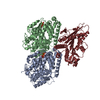
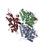
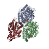
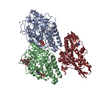
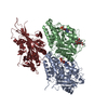
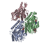
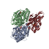
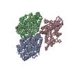
| Entry | Database: PDB / ID: 6mlr | ||||||
|---|---|---|---|---|---|---|---|
| Title | Cryo-EM structure of microtubule-bound Kif7 in the AMPPNP state | ||||||
 Components Components |
| ||||||
 Keywords Keywords | motor protein/inhibitor / Microtubule tip-tracking / Primary cilium / Hedgehog signaling / MOTOR PROTEIN / motor protein-inhibitor complex | ||||||
| Function / homology |  Function and homology information Function and homology informationpositive regulation of smoothened signaling pathway / microtubule motor activity / kinesin complex / microtubule-based movement / Hedgehog 'off' state / ciliary tip / negative regulation of smoothened signaling pathway / microtubule cytoskeleton organization / Hedgehog 'on' state / structural constituent of cytoskeleton ...positive regulation of smoothened signaling pathway / microtubule motor activity / kinesin complex / microtubule-based movement / Hedgehog 'off' state / ciliary tip / negative regulation of smoothened signaling pathway / microtubule cytoskeleton organization / Hedgehog 'on' state / structural constituent of cytoskeleton / fibrillar center / neuron migration / mitotic cell cycle / microtubule binding / microtubule / Hydrolases; Acting on acid anhydrides; Acting on GTP to facilitate cellular and subcellular movement / cilium / ciliary basal body / hydrolase activity / GTPase activity / GTP binding / ATP hydrolysis activity / ATP binding / metal ion binding / cytosol / cytoplasm Similarity search - Function | ||||||
| Biological species |  Homo sapiens (human) Homo sapiens (human) | ||||||
| Method | ELECTRON MICROSCOPY / helical reconstruction / cryo EM / Resolution: 4.2 Å | ||||||
 Authors Authors | Mani, N. / Jiang, S. / Wilson-Kubalek, E.M. / Ku, P. / Milligan, R.A. / Subramanian, R. | ||||||
| Funding support |  United States, 1items United States, 1items
| ||||||
 Citation Citation | Journal: Dev Cell / Year: 2019 Title: Interplay between the Kinesin and Tubulin Mechanochemical Cycles Underlies Microtubule Tip Tracking by the Non-motile Ciliary Kinesin Kif7. Authors: Shuo Jiang / Nandini Mani / Elizabeth M Wilson-Kubalek / Pei-I Ku / Ronald A Milligan / Radhika Subramanian / Abstract: The correct localization of Hedgehog effectors to the tip of primary cilia is critical for proper signal transduction. The conserved non-motile kinesin Kif7 defines a "cilium-tip compartment" by ...The correct localization of Hedgehog effectors to the tip of primary cilia is critical for proper signal transduction. The conserved non-motile kinesin Kif7 defines a "cilium-tip compartment" by localizing to the distal ends of axonemal microtubules. How Kif7 recognizes microtubule ends remains unknown. We find that Kif7 preferentially binds GTP-tubulin at microtubule ends over GDP-tubulin in the mature microtubule lattice, and ATP hydrolysis by Kif7 enhances this discrimination. Cryo-electron microscopy (cryo-EM) structures suggest that a rotated microtubule footprint and conformational changes in the ATP-binding pocket underlie Kif7's atypical microtubule-binding properties. Finally, Kif7 not only recognizes but also stabilizes a GTP-form of tubulin to promote its own microtubule-end localization. Thus, unlike the characteristic microtubule-regulated ATPase activity of kinesins, Kif7 modulates the tubulin mechanochemical cycle. We propose that the ubiquitous kinesin fold has been repurposed in Kif7 to facilitate organization of a spatially restricted platform for localization of Hedgehog effectors at the cilium tip. | ||||||
| History |
|
- Structure visualization
| Movie |
Movie viewer |
|---|---|
| Structure viewer | Molecule: MolmilJmol/JSmol |
- Downloads & links
-Download
| PDBx/mmCIF format | 6mlr.cif.gz | 223.9 KB | Display | PDBx/mmCIF format |
|---|---|---|---|---|
| PDB format | pdb6mlr.ent.gz | 171.9 KB | Display | PDB format |
| PDBx/mmJSON format | 6mlr.json.gz | Tree view | PDBx/mmJSON format | |
| Others |  Other downloads Other downloads |
-Validation report
| Arichive directory | https://data.pdbj.org/pub/pdb/validation_reports/ml/6mlrftp://data.pdbj.org/pub/pdb/validation_reports/ml/6mlr | HTTPS FTP |
|---|
-Related structure data
| Related structure data |  9141MC 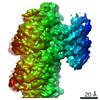 9140C 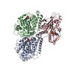 6mlqC M: map data used to model this data C: citing same article ( |
|---|---|
| Similar structure data |

-Links
 PDBj
PDBj


- Assembly
| Deposited unit | 
|
|---|---|
| 1 |
|
-Components
-Protein , 3 types, 3 molecules ABC
| #1: Protein | Mass: 50121.266 Da / Num. of mol.: 1 / Source method: isolated from a natural source / Source: (natural) |
|---|---|
| #2: Protein | Mass: 49907.770 Da / Num. of mol.: 1 / Source method: isolated from a natural source / Source: (natural) |
| #3: Protein | Mass: 43663.262 Da / Num. of mol.: 1 Source method: isolated from a genetically manipulated source Source: (gene. exp.) Homo sapiens (human) / Gene: KIF7, UNQ340/PRO539 / Plasmid: pET-28a(+) / Production host:  |
-Non-polymers , 4 types, 4 molecules 


| #4: Chemical | ChemComp-GTP /  Mass: 523.180 Da / Num. of mol.: 1 / Source method: obtained synthetically / Formula: C10H16N5O14P3 / Comment: GTP, energy-carrying molecule*YM Mass: 523.180 Da / Num. of mol.: 1 / Source method: obtained synthetically / Formula: C10H16N5O14P3 / Comment: GTP, energy-carrying molecule*YM |
|---|---|
| #5: Chemical | ChemComp-GDP /  Type: RNA linking / Mass: 443.201 Da / Num. of mol.: 1 / Source method: obtained synthetically / Formula: C10H15N5O11P2 / Comment: GDP, energy-carrying molecule*YM Type: RNA linking / Mass: 443.201 Da / Num. of mol.: 1 / Source method: obtained synthetically / Formula: C10H15N5O11P2 / Comment: GDP, energy-carrying molecule*YM |
| #6: Chemical | ChemComp-TA1 /  Mass: 853.906 Da / Num. of mol.: 1 / Source method: obtained synthetically / Formula: C47H51NO14 / Comment: medication, chemotherapy*YM Mass: 853.906 Da / Num. of mol.: 1 / Source method: obtained synthetically / Formula: C47H51NO14 / Comment: medication, chemotherapy*YM |
| #7: Chemical | ChemComp-ANP /  Mass: 506.196 Da / Num. of mol.: 1 / Source method: obtained synthetically / Formula: C10H17N6O12P3 / Comment: AMP-PNP, energy-carrying molecule analogue*YM Mass: 506.196 Da / Num. of mol.: 1 / Source method: obtained synthetically / Formula: C10H17N6O12P3 / Comment: AMP-PNP, energy-carrying molecule analogue*YM |
-Experimental details
-Experiment
| Experiment | Method: ELECTRON MICROSCOPY |
|---|---|
| EM experiment | Aggregation state: HELICAL ARRAY / 3D reconstruction method: helical reconstruction |
- Sample preparation
| Component | Name: Microtubule-bound Kif7 / Type: COMPLEX / Entity ID: #1-#3 / Source: RECOMBINANT |
|---|---|
| Source (natural) | Organism: |
| Source (recombinant) | Organism: |
| Buffer solution | pH: 6.8 |
| Specimen | Conc.: 0.5 mg/ml / Embedding applied: NO / Shadowing applied: NO / Staining applied: NO / Vitrification applied: YES |
| Specimen support | Details: not available |
| Vitrification | Instrument: HOMEMADE PLUNGER / Cryogen name: ETHANE / Humidity: 90 % / Chamber temperature: 280 K / Details: Blotted from behind the grid for 2 seconds |
- Electron microscopy imaging
| Experimental equipment |  Model: Titan Krios / Image courtesy: FEI Company |
|---|---|
| Microscopy | Model: FEI TITAN KRIOS |
| Electron gun | Electron source:  FIELD EMISSION GUN / Accelerating voltage: 300 kV / Illumination mode: FLOOD BEAM FIELD EMISSION GUN / Accelerating voltage: 300 kV / Illumination mode: FLOOD BEAM |
| Electron lens | Mode: BRIGHT FIELD |
| Image recording | Average exposure time: 8 sec. / Electron dose: 37 e/Å2 / Detector mode: COUNTING / Film or detector model: GATAN K2 SUMMIT (4k x 4k) / Num. of grids imaged: 1 / Num. of real images: 1059 |
| Image scans | Movie frames/image: 40 / Used frames/image: 0-40 |
- Processing
| Software | Name: PHENIX / Version: 1.12_2829: / Classification: refinement | ||||||||||||||||||||||||
|---|---|---|---|---|---|---|---|---|---|---|---|---|---|---|---|---|---|---|---|---|---|---|---|---|---|
| EM software |
| ||||||||||||||||||||||||
| Image processing | Details: The images were corrected by motionCorr2 | ||||||||||||||||||||||||
| CTF correction | Details: CTF correction using CTFFIND4 / Type: PHASE FLIPPING ONLY | ||||||||||||||||||||||||
| Helical symmerty | Angular rotation/subunit: -25.76 ° / Axial rise/subunit: 8.83 Å / Axial symmetry: C1 | ||||||||||||||||||||||||
| Particle selection | Num. of particles selected: 84000 Details: segmnets were picked along helical segments manually using appion | ||||||||||||||||||||||||
| 3D reconstruction | Resolution: 4.2 Å / Resolution method: FSC 0.143 CUT-OFF / Num. of particles: 37220 / Algorithm: BACK PROJECTION / Num. of class averages: 1 / Symmetry type: HELICAL | ||||||||||||||||||||||||
| Atomic model building | Protocol: FLEXIBLE FIT | ||||||||||||||||||||||||
| Atomic model building | 3D fitting-ID: 1 / Source name: PDB / Type: experimental model
| ||||||||||||||||||||||||
| Refinement | Highest resolution: 4.2 Å | ||||||||||||||||||||||||
| Refine LS restraints |
|