Movie
Movie Controller


+ Open data

- Basic information
| Entry | Database: EMDB / ID: EMD-9141 | |||||||||
|---|---|---|---|---|---|---|---|---|---|---|
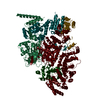
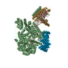
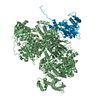
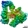
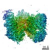
| Title | Cryo-EM structure of microtubule-bound Kif7 in the AMPPNP state | |||||||||
 Map data Map data | Kif7 bound to microtubules in the AMPPNP state | |||||||||
 Sample Sample |
| |||||||||
 Keywords Keywords | Microtubule tip-tracking / Primary cilium / Hedgehog signaling / MOTOR PROTEIN / motor protein-inhibitor complex | |||||||||
| Function / homology |  Function and homology information Function and homology informationpositive regulation of smoothened signaling pathway / microtubule motor activity / kinesin complex / microtubule-based movement / Hedgehog 'off' state / ciliary tip / negative regulation of smoothened signaling pathway / microtubule cytoskeleton organization / Hedgehog 'on' state / structural constituent of cytoskeleton ...positive regulation of smoothened signaling pathway / microtubule motor activity / kinesin complex / microtubule-based movement / Hedgehog 'off' state / ciliary tip / negative regulation of smoothened signaling pathway / microtubule cytoskeleton organization / Hedgehog 'on' state / structural constituent of cytoskeleton / fibrillar center / neuron migration / mitotic cell cycle / microtubule binding / microtubule / Hydrolases; Acting on acid anhydrides; Acting on GTP to facilitate cellular and subcellular movement / cilium / ciliary basal body / hydrolase activity / GTPase activity / GTP binding / ATP hydrolysis activity / ATP binding / metal ion binding / cytosol / cytoplasm Similarity search - Function | |||||||||
| Biological species |   Homo sapiens (human) Homo sapiens (human) | |||||||||
| Method | helical reconstruction / cryo EM / Resolution: 4.2 Å | |||||||||
 Authors Authors | Wilson-Kubalek EM / Jiang S / Mani N / Ku P / Milligan RA / Subramanian R | |||||||||
| Funding support |  United States, 1 items United States, 1 items
| |||||||||
 Citation Citation | Journal: Dev Cell / Year: 2019 Title: Interplay between the Kinesin and Tubulin Mechanochemical Cycles Underlies Microtubule Tip Tracking by the Non-motile Ciliary Kinesin Kif7. Authors: Shuo Jiang / Nandini Mani / Elizabeth M Wilson-Kubalek / Pei-I Ku / Ronald A Milligan / Radhika Subramanian / Abstract: The correct localization of Hedgehog effectors to the tip of primary cilia is critical for proper signal transduction. The conserved non-motile kinesin Kif7 defines a "cilium-tip compartment" by ...The correct localization of Hedgehog effectors to the tip of primary cilia is critical for proper signal transduction. The conserved non-motile kinesin Kif7 defines a "cilium-tip compartment" by localizing to the distal ends of axonemal microtubules. How Kif7 recognizes microtubule ends remains unknown. We find that Kif7 preferentially binds GTP-tubulin at microtubule ends over GDP-tubulin in the mature microtubule lattice, and ATP hydrolysis by Kif7 enhances this discrimination. Cryo-electron microscopy (cryo-EM) structures suggest that a rotated microtubule footprint and conformational changes in the ATP-binding pocket underlie Kif7's atypical microtubule-binding properties. Finally, Kif7 not only recognizes but also stabilizes a GTP-form of tubulin to promote its own microtubule-end localization. Thus, unlike the characteristic microtubule-regulated ATPase activity of kinesins, Kif7 modulates the tubulin mechanochemical cycle. We propose that the ubiquitous kinesin fold has been repurposed in Kif7 to facilitate organization of a spatially restricted platform for localization of Hedgehog effectors at the cilium tip. | |||||||||
| History |
|
- Structure visualization
| Movie |
Movie viewer |
|---|---|
| Structure viewer | EM map: SurfViewMolmilJmol/JSmol |
| Supplemental images |

- Downloads & links
-EMDB archive
| Map data | emd_9141.map.gz | 2.4 MB | EMDB map data format | |
|---|---|---|---|---|
| Header (meta data) | emd-9141-v30.xmlemd-9141.xml | 16.4 KB 16.4 KB | Display Display | EMDB header |
| Images | 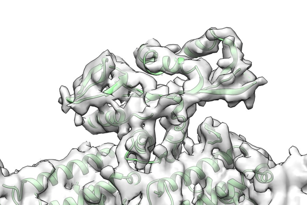 emd_9141.png emd_9141.png | 268.5 KB | ||
| Filedesc metadata | emd-9141.cif.gz | 7.3 KB | ||
| Archive directory |  http://ftp.pdbj.org/pub/emdb/structures/EMD-9141ftp://ftp.pdbj.org/pub/emdb/structures/EMD-9141 http://ftp.pdbj.org/pub/emdb/structures/EMD-9141ftp://ftp.pdbj.org/pub/emdb/structures/EMD-9141 | HTTPS FTP |
-Related structure data
| Related structure data |  6mlrMC 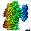 9140C 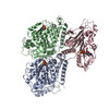 6mlqC M: atomic model generated by this map C: citing same article ( |
|---|---|
| Similar structure data |
-Links
| EMDB pages | EMDB (EBI/PDBe) / EMDataResource |
|---|---|
| Related items in Molecule of the Month |


-Map
| File | Download / File: emd_9141.map.gz / Format: CCP4 / Size: 2.6 MB / Type: IMAGE STORED AS FLOATING POINT NUMBER (4 BYTES) | ||||||||||||||||||||||||||||||||||||||||||||||||||||||||||||
|---|---|---|---|---|---|---|---|---|---|---|---|---|---|---|---|---|---|---|---|---|---|---|---|---|---|---|---|---|---|---|---|---|---|---|---|---|---|---|---|---|---|---|---|---|---|---|---|---|---|---|---|---|---|---|---|---|---|---|---|---|---|
| Annotation | Kif7 bound to microtubules in the AMPPNP state | ||||||||||||||||||||||||||||||||||||||||||||||||||||||||||||
| Projections & slices | Image control
Images are generated by Spider. generated in cubic-lattice coordinate | ||||||||||||||||||||||||||||||||||||||||||||||||||||||||||||
| Voxel size | X=Y=Z: 1.31 Å | ||||||||||||||||||||||||||||||||||||||||||||||||||||||||||||
| Density |
| ||||||||||||||||||||||||||||||||||||||||||||||||||||||||||||
| Symmetry | Space group: 1 | ||||||||||||||||||||||||||||||||||||||||||||||||||||||||||||
| Details | EMDB XML:
CCP4 map header:
| ||||||||||||||||||||||||||||||||||||||||||||||||||||||||||||
 Z (Sec.)
 Y (Row.)
 X (Col.)


-Supplemental data
- Sample components
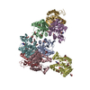
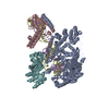
-Entire : Microtubule-bound Kif7
| Entire | Name: Microtubule-bound Kif7 |
|---|---|
| Components |
|
-Supramolecule #1: Microtubule-bound Kif7
| Supramolecule | Name: Microtubule-bound Kif7 / type: complex / ID: 1 / Parent: 0 / Macromolecule list: #1-#3 |
|---|---|
| Source (natural) | Organism: |
-Macromolecule #1: Tubulin alpha-1A chain
| Macromolecule | Name: Tubulin alpha-1A chain / type: protein_or_peptide / ID: 1 / Number of copies: 1 / Enantiomer: LEVO |
|---|---|
| Source (natural) | Organism: |
| Molecular weight | Theoretical: 50.121266 KDa |
| Sequence | String: MRECISIHVG QAGVQIGNAC WELYCLEHGI QPDGQMPSDK TIGGGDDSFN TFFSETGAGK HVPRAVFVDL EPTVIDEVRT GTYRQLFHP EQLITGKEDA ANNYARGHYT IGKEIIDLVL DRIRKLADQC TGLQGFSVFH SFGGGTGSGF TSLLMERLSV D YGKKSKLE ...String: MRECISIHVG QAGVQIGNAC WELYCLEHGI QPDGQMPSDK TIGGGDDSFN TFFSETGAGK HVPRAVFVDL EPTVIDEVRT GTYRQLFHP EQLITGKEDA ANNYARGHYT IGKEIIDLVL DRIRKLADQC TGLQGFSVFH SFGGGTGSGF TSLLMERLSV D YGKKSKLE FSIYPAPQVS TAVVEPYNSI LTTHTTLEHS DCAFMVDNEA IYDICRRNLD IERPTYTNLN RLIGQIVSSI TA SLRFDGA LNVDLTEFQT NLVPYPRAHF PLATYAPVIS AEKAYHEQLS VAEITNACFE PANQMVKCDP RHGKYMACCL LYR GDVVPK DVNAAIATIK TKRTIQFVDW CPTGFKVGIN YEPPTVVPGG DLAKVQRAVC MLSNTTAIAE AWARLDHKFD LMYA KRAFV HWYVGEGMEE GEFSEAREDM AALEKDYEEV GVDSVEGEGE EEGEEY UniProtKB: Tubulin alpha-1A chain |
-Macromolecule #2: Tubulin beta chain
| Macromolecule | Name: Tubulin beta chain / type: protein_or_peptide / ID: 2 / Number of copies: 1 / Enantiomer: LEVO |
|---|---|
| Source (natural) | Organism: |
| Molecular weight | Theoretical: 49.90777 KDa |
| Sequence | String: MREIVHIQAG QCGNQIGAKF WEVISDEHGI DPTGSYHGDS DLQLERINVY YNEAAGNKYV PRAILVDLEP GTMDSVRSGP FGQIFRPDN FVFGQSGAGN NWAKGHYTEG AELVDSVLDV VRKESESCDC LQGFQLTHSL GGGTGSGMGT LLISKIREEY P DRIMNTFS ...String: MREIVHIQAG QCGNQIGAKF WEVISDEHGI DPTGSYHGDS DLQLERINVY YNEAAGNKYV PRAILVDLEP GTMDSVRSGP FGQIFRPDN FVFGQSGAGN NWAKGHYTEG AELVDSVLDV VRKESESCDC LQGFQLTHSL GGGTGSGMGT LLISKIREEY P DRIMNTFS VVPSPKVSDT VVEPYNATLS VHQLVENTDE TYCIDNEALY DICFRTLKLT TPTYGDLNHL VSATMSGVTT CL RFPGQLN ADLRKLAVNM VPFPRLHFFM PGFAPLTSRG SQQYRALTVP ELTQQMFDAK NMMAACDPRH GRYLTVAAVF RGR MSMKEV DEQMLNVQNK NSSYFVEWIP NNVKTAVCDI PPRGLKMSAT FIGNSTAIQE LFKRISEQFT AMFRRKAFLH WYTG EGMDE MEFTEAESNM NDLVSEYQQY QDATADEQGE FEEEGEEDEA UniProtKB: Tubulin beta chain |
-Macromolecule #3: Kinesin-like protein KIF7
| Macromolecule | Name: Kinesin-like protein KIF7 / type: protein_or_peptide / ID: 3 / Number of copies: 1 / Enantiomer: LEVO |
|---|---|
| Source (natural) | Organism: Homo sapiens (human) |
| Molecular weight | Theoretical: 43.663262 KDa |
| Recombinant expression | Organism:  |
| Sequence | String: GMGLEAQRLP GAEEAPVRVA LRVRPLLPKE LLHGHQSCLQ VEPGLGRVTL GRDRHFGFHV VLAEDAGQEA VYQACVQPLL EAFFEGFNA TVFAYGQTGS GKTYTMGEAS VASLLEDEQG IVPRAMAEAF KLIDENDLLD CLVHVSYLEV YKEEFRDLLE V GTASRDIQ ...String: GMGLEAQRLP GAEEAPVRVA LRVRPLLPKE LLHGHQSCLQ VEPGLGRVTL GRDRHFGFHV VLAEDAGQEA VYQACVQPLL EAFFEGFNA TVFAYGQTGS GKTYTMGEAS VASLLEDEQG IVPRAMAEAF KLIDENDLLD CLVHVSYLEV YKEEFRDLLE V GTASRDIQ LREDERGNVV LCGVKEVDVE GLDEVLSLLE MGNAARHTGA THLNHLSSRS HTVFTVTLEQ RGRAPSRLPR PA PGQLLVS KFHFVDLAGS ERVLKTGSTG ERLKESIQIN SSLLALGNVI SALGDPQRRG SHIPYRDSKI TRILKDSLGG NAK TVMIAC VSPSSSDFDE TLNTLNYASR AQNIRNRATV NWRPEAERPP EETASGARGP PRHRSETRII HRGRRAPGPA TAS UniProtKB: Kinesin-like protein KIF7 |
-Macromolecule #4: GUANOSINE-5'-TRIPHOSPHATE
| Macromolecule | Name: GUANOSINE-5'-TRIPHOSPHATE / type: ligand / ID: 4 / Number of copies: 1 / Formula: GTP |
|---|---|
| Molecular weight | Theoretical: 523.18 Da |
| Chemical component information |  ChemComp-GTP: |
-Macromolecule #5: GUANOSINE-5'-DIPHOSPHATE
| Macromolecule | Name: GUANOSINE-5'-DIPHOSPHATE / type: ligand / ID: 5 / Number of copies: 1 / Formula: GDP |
|---|---|
| Molecular weight | Theoretical: 443.201 Da |
| Chemical component information |  ChemComp-GDP: |
-Macromolecule #6: TAXOL
| Macromolecule | Name: TAXOL / type: ligand / ID: 6 / Number of copies: 1 / Formula: TA1 |
|---|---|
| Molecular weight | Theoretical: 853.906 Da |
| Chemical component information |  ChemComp-TA1: |
-Macromolecule #7: PHOSPHOAMINOPHOSPHONIC ACID-ADENYLATE ESTER
| Macromolecule | Name: PHOSPHOAMINOPHOSPHONIC ACID-ADENYLATE ESTER / type: ligand / ID: 7 / Number of copies: 1 / Formula: ANP |
|---|---|
| Molecular weight | Theoretical: 506.196 Da |
| Chemical component information |  ChemComp-ANP: |
-Experimental details
-Structure determination
| Method | cryo EM |
|---|---|
 Processing Processing | helical reconstruction |
| Aggregation state | helical array |
-Sample preparation
| Concentration | 0.5 mg/mL |
|---|---|
| Buffer | pH: 6.8 |
| Grid | Support film - Material: CARBON / Support film - topology: HOLEY / Details: not available |
| Vitrification | Cryogen name: ETHANE / Chamber humidity: 90 % / Chamber temperature: 280 K / Instrument: HOMEMADE PLUNGER / Details: Blotted from behind the grid for 2 seconds. |
- Electron microscopy
| Microscope | FEI TITAN KRIOS |
|---|---|
| Image recording | Film or detector model: GATAN K2 SUMMIT (4k x 4k) / Detector mode: COUNTING / Digitization - Frames/image: 0-40 / Number grids imaged: 1 / Number real images: 1059 / Average exposure time: 8.0 sec. / Average electron dose: 37.0 e/Å2 |
| Electron beam | Acceleration voltage: 300 kV / Electron source:  FIELD EMISSION GUN FIELD EMISSION GUN |
| Electron optics | Illumination mode: FLOOD BEAM / Imaging mode: BRIGHT FIELD |
| Experimental equipment |  Model: Titan Krios / Image courtesy: FEI Company |
+Image processing
-Atomic model buiding 1
| Initial model |
| ||||||||
|---|---|---|---|---|---|---|---|---|---|
| Refinement | Protocol: FLEXIBLE FIT | ||||||||
| Output model | PDB-6mlr: |