 Movie
Movie Controller


+ Open data

- Basic information
| Entry | Database: PDB / ID: 6lhu | |||||||||||||||||||||||||||
|---|---|---|---|---|---|---|---|---|---|---|---|---|---|---|---|---|---|---|---|---|---|---|---|---|---|---|---|---|
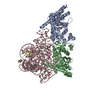
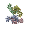
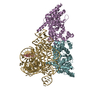
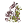
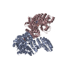
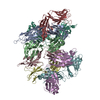
| Title | High resolution structure of FANCA C-terminal domain (CTD) | |||||||||||||||||||||||||||
 Components Components | Fanconi anemia complementation group A | |||||||||||||||||||||||||||
 Keywords Keywords | DNA REPAIR / nuclear localization / fanconi anemia core protein / fanconi anemia complementation group a / interstrand crosslink repair | |||||||||||||||||||||||||||
| Function / homology |  Function and homology information Function and homology information | |||||||||||||||||||||||||||
| Biological species | ||||||||||||||||||||||||||||
| Method | ELECTRON MICROSCOPY / single particle reconstruction / cryo EM / Resolution: 3.46 Å | |||||||||||||||||||||||||||
 Authors Authors | Jeong, E. / Lee, S. / Shin, J. / Kim, Y. / Kim, J. / Scharer, O. / Kim, Y. / Kim, H. / Cho, Y. | |||||||||||||||||||||||||||
| Funding support |  Korea, Republic Of, 2items Korea, Republic Of, 2items
| |||||||||||||||||||||||||||
 Citation Citation | Journal: Nucleic Acids Res / Year: 2020 Title: Structural basis of the fanconi anemia-associated mutations within the FANCA and FANCG complex. Authors: Eunyoung Jeong / Seong-Gyu Lee / Hyun-Suk Kim / Jihyeon Yang / Jinwoo Shin / Youngran Kim / Jihan Kim / Orlando D Schärer / Youngjin Kim / Jung-Eun Yeo / Ho Min Kim / Yunje Cho / Abstract: Monoubiquitination of the Fanconi anemia complementation group D2 (FANCD2) protein by the FA core ubiquitin ligase complex is the central event in the FA pathway. FANCA and FANCG play major roles in ...Monoubiquitination of the Fanconi anemia complementation group D2 (FANCD2) protein by the FA core ubiquitin ligase complex is the central event in the FA pathway. FANCA and FANCG play major roles in the nuclear localization of the FA core complex. Mutations of these two genes are the most frequently observed genetic alterations in FA patients, and most point mutations in FANCA are clustered in the C-terminal domain (CTD). To understand the basis of the FA-associated FANCA mutations, we determined the cryo-electron microscopy (EM) structures of Xenopus laevis FANCA alone at 3.35 Å and 3.46 Å resolution and two distinct FANCA-FANCG complexes at 4.59 and 4.84 Å resolution, respectively. The FANCA CTD adopts an arc-shaped solenoid structure that forms a pseudo-symmetric dimer through its outer surface. FA- and cancer-associated point mutations are widely distributed over the CTD. The two different complex structures capture independent interactions of FANCG with either FANCA C-terminal HEAT repeats, or the N-terminal region. We show that mutations that disturb either of these two interactions prevent the nuclear localization of FANCA, thereby leading to an FA pathway defect. The structure provides insights into the function of FANCA CTD, and provides a framework for understanding FA- and cancer-associated mutations. | |||||||||||||||||||||||||||
| History |
|
- Structure visualization
| Movie |
Movie viewer |
|---|---|
| Structure viewer | Molecule: MolmilJmol/JSmol |
- Downloads & links
-Download
| PDBx/mmCIF format | 6lhu.cif.gz | 510.6 KB | Display | PDBx/mmCIF format |
|---|---|---|---|---|
| PDB format | pdb6lhu.ent.gz | 391.8 KB | Display | PDB format |
| PDBx/mmJSON format | 6lhu.json.gz | Tree view | PDBx/mmJSON format | |
| Others |  Other downloads Other downloads |
-Validation report
| Arichive directory | https://data.pdbj.org/pub/pdb/validation_reports/lh/6lhuftp://data.pdbj.org/pub/pdb/validation_reports/lh/6lhu | HTTPS FTP |
|---|
-Related structure data
| Related structure data | 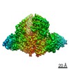 0899MC 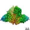 0896C 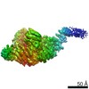 0900C 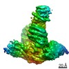 0901C 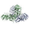 6lhsC  6lhvC 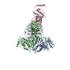 6lhwC M: map data used to model this data C: citing same article ( |
|---|---|
| Similar structure data |
-Links
 PDBj
PDBj- Assembly
| Deposited unit | 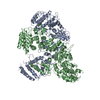
|
|---|---|
| 1 |
|
-Components
| #1: Protein | Mass: 163009.344 Da / Num. of mol.: 2 Source method: isolated from a genetically manipulated source Source: (gene. exp.)  Trichoplusia ni (cabbage looper) / References: UniProt: Q4VT51 Trichoplusia ni (cabbage looper) / References: UniProt: Q4VT51Has protein modification | N | |
|---|
-Experimental details
-Experiment
| Experiment | Method: ELECTRON MICROSCOPY |
|---|---|
| EM experiment | Aggregation state: PARTICLE / 3D reconstruction method: single particle reconstruction |
- Sample preparation
| Component | Name: FANCA / Type: COMPLEX / Details: homo dimer / Entity ID: all / Source: RECOMBINANT |
|---|---|
| Molecular weight | Value: 500 kDa/nm / Experimental value: YES |
| Source (natural) | Organism: |
| Source (recombinant) | Organism: Trichoplusia ni (cabbage looper) |
| Buffer solution | pH: 8 |
| Specimen | Conc.: 0.8 mg/ml / Embedding applied: NO / Shadowing applied: NO / Staining applied: NO / Vitrification applied: YES |
| Vitrification | Cryogen name: ETHANE |
- Electron microscopy imaging
| Experimental equipment |  Model: Titan Krios / Image courtesy: FEI Company |
|---|---|
| Microscopy | Model: FEI TITAN KRIOS |
| Electron gun | Electron source:  FIELD EMISSION GUN / Accelerating voltage: 300 kV / Illumination mode: FLOOD BEAM FIELD EMISSION GUN / Accelerating voltage: 300 kV / Illumination mode: FLOOD BEAM |
| Electron lens | Mode: DIFFRACTION |
| Image recording | Electron dose: 30 e/Å2 / Film or detector model: FEI FALCON III (4k x 4k) |
- Processing
| Software | Name: PHENIX / Version: 1.15.2_3472: / Classification: refinement | ||||||||||||||||||||||||
|---|---|---|---|---|---|---|---|---|---|---|---|---|---|---|---|---|---|---|---|---|---|---|---|---|---|
| EM software | Name: PHENIX / Category: model refinement | ||||||||||||||||||||||||
| CTF correction | Type: PHASE FLIPPING AND AMPLITUDE CORRECTION | ||||||||||||||||||||||||
| 3D reconstruction | Resolution: 3.46 Å / Resolution method: FSC 0.143 CUT-OFF / Num. of particles: 150141 / Symmetry type: POINT | ||||||||||||||||||||||||
| Refine LS restraints |
|
