 Movie
Movie Controller


+ Open data

- Basic information
| Entry | Database: EMDB / ID: EMD-0899 | |||||||||
|---|---|---|---|---|---|---|---|---|---|---|
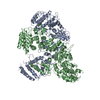
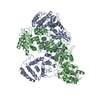
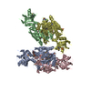
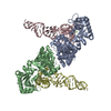
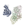
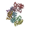
| Title | High resolution structure of FANCA C-terminal domain (CTD) | |||||||||
 Map data Map data | ||||||||||
 Sample Sample |
| |||||||||
 Keywords Keywords | nuclear localization / fanconi anemia core protein / fanconi anemia complementation group a / interstrand crosslink repair / DNA REPAIR | |||||||||
| Function / homology |  Function and homology information Function and homology information | |||||||||
| Biological species | ||||||||||
| Method | single particle reconstruction / cryo EM / Resolution: 3.46 Å | |||||||||
 Authors Authors | Jeong E / Lee S | |||||||||
| Funding support |  Korea, Republic Of, 2 items Korea, Republic Of, 2 items
| |||||||||
 Citation Citation | Journal: Nucleic Acids Res / Year: 2020 Title: Structural basis of the fanconi anemia-associated mutations within the FANCA and FANCG complex. Authors: Eunyoung Jeong / Seong-Gyu Lee / Hyun-Suk Kim / Jihyeon Yang / Jinwoo Shin / Youngran Kim / Jihan Kim / Orlando D Schärer / Youngjin Kim / Jung-Eun Yeo / Ho Min Kim / Yunje Cho / Abstract: Monoubiquitination of the Fanconi anemia complementation group D2 (FANCD2) protein by the FA core ubiquitin ligase complex is the central event in the FA pathway. FANCA and FANCG play major roles in ...Monoubiquitination of the Fanconi anemia complementation group D2 (FANCD2) protein by the FA core ubiquitin ligase complex is the central event in the FA pathway. FANCA and FANCG play major roles in the nuclear localization of the FA core complex. Mutations of these two genes are the most frequently observed genetic alterations in FA patients, and most point mutations in FANCA are clustered in the C-terminal domain (CTD). To understand the basis of the FA-associated FANCA mutations, we determined the cryo-electron microscopy (EM) structures of Xenopus laevis FANCA alone at 3.35 Å and 3.46 Å resolution and two distinct FANCA-FANCG complexes at 4.59 and 4.84 Å resolution, respectively. The FANCA CTD adopts an arc-shaped solenoid structure that forms a pseudo-symmetric dimer through its outer surface. FA- and cancer-associated point mutations are widely distributed over the CTD. The two different complex structures capture independent interactions of FANCG with either FANCA C-terminal HEAT repeats, or the N-terminal region. We show that mutations that disturb either of these two interactions prevent the nuclear localization of FANCA, thereby leading to an FA pathway defect. The structure provides insights into the function of FANCA CTD, and provides a framework for understanding FA- and cancer-associated mutations. | |||||||||
| History |
|
- Structure visualization
| Movie |
Movie viewer |
|---|---|
| Structure viewer | EM map: SurfViewMolmilJmol/JSmol |
| Supplemental images |

- Downloads & links
-EMDB archive
| Map data | emd_0899.map.gz | 59.7 MB | EMDB map data format | |
|---|---|---|---|---|
| Header (meta data) | emd-0899-v30.xmlemd-0899.xml | 13.9 KB 13.9 KB | Display Display | EMDB header |
| Images | 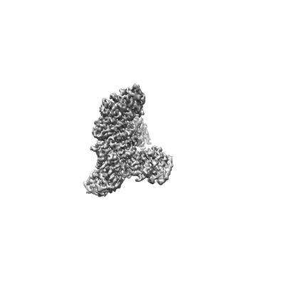 emd_0899.png emd_0899.png | 13.7 KB | ||
| Filedesc metadata | emd-0899.cif.gz | 6.4 KB | ||
| Archive directory |  http://ftp.pdbj.org/pub/emdb/structures/EMD-0899ftp://ftp.pdbj.org/pub/emdb/structures/EMD-0899 http://ftp.pdbj.org/pub/emdb/structures/EMD-0899ftp://ftp.pdbj.org/pub/emdb/structures/EMD-0899 | HTTPS FTP |
-Related structure data
| Related structure data |  6lhuMC 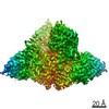 0896C 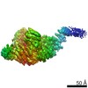 0900C 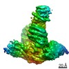 0901C 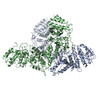 6lhsC 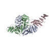 6lhvC 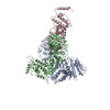 6lhwC C: citing same article ( M: atomic model generated by this map |
|---|---|
| Similar structure data |

-Links
| EMDB pages | EMDB (EBI/PDBe) / EMDataResource |
|---|
-Map
| File | Download / File: emd_0899.map.gz / Format: CCP4 / Size: 64 MB / Type: IMAGE STORED AS FLOATING POINT NUMBER (4 BYTES) | ||||||||||||||||||||||||||||||||||||||||||||||||||||||||||||
|---|---|---|---|---|---|---|---|---|---|---|---|---|---|---|---|---|---|---|---|---|---|---|---|---|---|---|---|---|---|---|---|---|---|---|---|---|---|---|---|---|---|---|---|---|---|---|---|---|---|---|---|---|---|---|---|---|---|---|---|---|---|
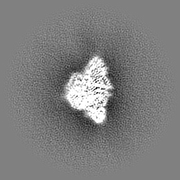
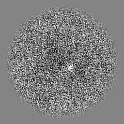
| Projections & slices | Image control
Images are generated by Spider. | ||||||||||||||||||||||||||||||||||||||||||||||||||||||||||||
| Voxel size | X=Y=Z: 1.4 Å | ||||||||||||||||||||||||||||||||||||||||||||||||||||||||||||
| Density |
| ||||||||||||||||||||||||||||||||||||||||||||||||||||||||||||
| Symmetry | Space group: 1 | ||||||||||||||||||||||||||||||||||||||||||||||||||||||||||||
| Details | EMDB XML:
CCP4 map header:
| ||||||||||||||||||||||||||||||||||||||||||||||||||||||||||||
 Z (Sec.)
 Y (Row.)
 X (Col.)


-Supplemental data
- Sample components
-Entire : FANCA
| Entire | Name: FANCA |
|---|---|
| Components |
|
-Supramolecule #1: FANCA
| Supramolecule | Name: FANCA / type: complex / ID: 1 / Parent: 0 / Macromolecule list: all / Details: homo dimer |
|---|---|
| Source (natural) | Organism: |
| Molecular weight | Theoretical: 500 kDa/nm |
-Macromolecule #1: Fanconi anemia complementation group A
| Macromolecule | Name: Fanconi anemia complementation group A / type: protein_or_peptide / ID: 1 / Number of copies: 2 / Enantiomer: LEVO |
|---|---|
| Source (natural) | Organism: |
| Molecular weight | Theoretical: 163.009344 KDa |
| Recombinant expression | Organism:  Trichoplusia ni (cabbage looper) Trichoplusia ni (cabbage looper) |
| Sequence | String: MWSHPQFEKG SGGSGMSAVS GFTPSGQKRS LAELLDGRVK RLDRKSNNSV LQEAALYLLS CNQDVSQFLS EVEAPPYKKT CNPENPVSI KSRMPSAAFV GSTLKDQASC LKISPGILTA KAAVANIQQI CQACGDSSAV LNPEQREKLC SLLKTLKILL A ENCFCRSL ...String: MWSHPQFEKG SGGSGMSAVS GFTPSGQKRS LAELLDGRVK RLDRKSNNSV LQEAALYLLS CNQDVSQFLS EVEAPPYKKT CNPENPVSI KSRMPSAAFV GSTLKDQASC LKISPGILTA KAAVANIQQI CQACGDSSAV LNPEQREKLC SLLKTLKILL A ENCFCRSL FCKEIWIHRP PLVFEAVWHL HNEGIVCLDE ILESCKDTIS AVDWLFSEMC SLCLYIDNSS LAGDLAEKMI SD FQALLVE NSFRRSSATE RILEQHKTEE ICLSILDKLL SWLLDAVSVE KSDKSSAEQH WLSAFEVHRY RARVVPESIE QFF IHTLTQ VLTFKPKLKV SDAIQCQANW SFVKTSTLLT DLYRKLFVAL SAEKLIAHIQ LVLDTQEVNW HHVLTCVSCL VICL PEAQQ LIKDLLCRLL THAFESYELE GMITAFLIVR QAALEGPAAF VSYTEWFKCT FGAANSYHGN SKKSLVFLLK FLSDI VPFE APQYLKVHVL HPPFVPTKYR PLLMEYISLA KTRLTDMKVS IEDMGLYEDL SARSNKVQPE SQAHQDVEKA LNIFEN TGK IPASVMEASI FRRPYFTSRF LPALLTPRVL PAAPDALMLL IDSMKRADKI PTNMFNAYIE ACEQEKLRKQ KGRQQMD QS LPDEPLGILQ SALSDLRPLV TDANKYEDVS AQVAVISEKL IAVMGEQKVD DDQVAAKFLK LEDGAQLDIQ EQTVADLL L TCFCQCLIAA SGTNPPDRQG QWPTLYVKML CGHQWAFAAV LRRMLQLLRF QAPFLKDSHI VGLAAFSIHL HECQPSLQF LITGVQNLEH YWENLLNLLC SDSVGVCLKL CTAAISYAFC RFSELHQDIF SGCVPPLFLR KLQYLVPRLI WETRGEVIRD DEEADSPLN WNLYALAGWK EAALSLWNQN RLQGLLREKS FQVTFMDWLL WEMTLKSNND VLCDTDRQEY QRWAVNHYLS E SSVVGGCN GDLERGCITI AEAVLQFSNR HIQHSEWESR NISMLKSHTG LGDILCRLQE LICDIVTSHH QKGRRHFFFA IF YQRLELH KGKKELSNHL SKQGVLEMCC RILLGLPPLF LINTPSEKGI RTLGSEDFWQ FVNKELKNLG PRGYALPYNI TAH FFRGVI SASVQCKDSS EAVNSILSAT YSTCPALLIS AAVGWPQLDP VLRSQWCSLF GVDLPKELRT LREQQASVDS CLSQ GEKLS LSCTPWLSAA FLYSTVQRKK LPCSRMLEIL DGLSSNFSMV LISLLFFSVM DIIYMFLKDG RKHKDLLENC VHIIH CLEQ KGETWVWLFQ MTDERKPELG LHLHRAASDV FLNLMPFAFF WLVPSLQLEQ VVQQQDFLVI ALDMYHKFLQ LFVDGS PLS SLSAKSHHLD SHDVFTCGRQ FLLCCVPKCQ KPNSAILKKM LESWEEHDPE LAAVLTRSFK APDDYDDLFF EPVF UniProtKB: FA complementation group A L homeolog |
-Experimental details
-Structure determination
| Method | cryo EM |
|---|---|
 Processing Processing | single particle reconstruction |
| Aggregation state | particle |
-Sample preparation
| Concentration | 0.8 mg/mL |
|---|---|
| Buffer | pH: 8 |
| Vitrification | Cryogen name: ETHANE |
- Electron microscopy
| Microscope | FEI TITAN KRIOS |
|---|---|
| Image recording | Film or detector model: FEI FALCON III (4k x 4k) / Average electron dose: 30.0 e/Å2 |
| Electron beam | Acceleration voltage: 300 kV / Electron source:  FIELD EMISSION GUN FIELD EMISSION GUN |
| Electron optics | Illumination mode: FLOOD BEAM / Imaging mode: DIFFRACTION |
| Experimental equipment |  Model: Titan Krios / Image courtesy: FEI Company |
