Movie
Movie Controller

[English] 日本語
 Yorodumi
- PDB-6iug: Cryo-EM structure of the plant actin filaments from Zea mays pollen -
+ Open data

- Basic information
| Entry | Database: PDB / ID: 6iug | ||||||||||||||||||
|---|---|---|---|---|---|---|---|---|---|---|---|---|---|---|---|---|---|---|---|
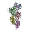
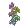
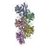
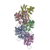
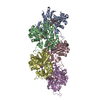
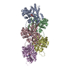
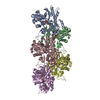
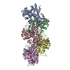
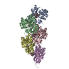
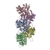
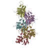
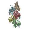
| Title | Cryo-EM structure of the plant actin filaments from Zea mays pollen | ||||||||||||||||||
 Components Components | pollen F-actin | ||||||||||||||||||
 Keywords Keywords | PROTEIN FIBRIL / Microfilament / Helix / Actin | ||||||||||||||||||
| Function / homology |  Function and homology information Function and homology information | ||||||||||||||||||
| Biological species |  | ||||||||||||||||||
| Method | ELECTRON MICROSCOPY / helical reconstruction / cryo EM / Resolution: 3.9 Å | ||||||||||||||||||
 Authors Authors | Ren, Z.H. / Zhang, Y. / Zhang, Y. / He, Y.Q. / Du, P.Z. / Wang, Z.X. / Sun, F. / Ren, H.Y. | ||||||||||||||||||
| Funding support |  China, 5items China, 5items
| ||||||||||||||||||
 Citation Citation | Journal: Plant Cell / Year: 2019 Title: Cryo-EM Structure of Actin Filaments from Pollen. Authors: Zhanhong Ren / Yan Zhang / Yi Zhang / Yunqiu He / Pingzhou Du / Zhanxin Wang / Fei Sun / Haiyun Ren / Abstract: Actins are among the most abundant and conserved proteins in eukaryotic cells, where they form filamentous structures that perform vital roles in key cellular processes. Although large amounts of ...Actins are among the most abundant and conserved proteins in eukaryotic cells, where they form filamentous structures that perform vital roles in key cellular processes. Although large amounts of data on the biochemical activities, dynamic behaviors, and important cellular functions of plant actin filaments have accumulated, their structural basis remains elusive. Here, we report a 3.9 Å structure of the plant actin filament from pollen (ZMPA) using cryo-electron microscopy. The structure shows a right-handed, double-stranded (two parallel strands) and staggered architecture that is stabilized by intra- and interstrand interactions. While the overall structure resembles that of other actin filaments, its DNase I binding loop bends farther outward, adopting an open conformation similar to that of the jasplakinolide- or beryllium fluoride (BeF)-stabilized rabbit skeletal muscle actin (RSMA) filament. Single-molecule magnetic tweezers analysis revealed that the ZMPA filament can resist a greater stretching force than the RSMA filament. Overall, these data provide evidence that plant actin filaments have greater stability than animal actin filaments, which might be important to their role as tracks for long-distance vesicle and organelle transportation.plantcell;31/12/2855/FX1F1fx1. | ||||||||||||||||||
| History |
|
- Structure visualization
| Movie |
Movie viewer |
|---|---|
| Structure viewer | Molecule: MolmilJmol/JSmol |
- Downloads & links
-Download
| PDBx/mmCIF format | 6iug.cif.gz | 325.8 KB | Display | PDBx/mmCIF format |
|---|---|---|---|---|
| PDB format | pdb6iug.ent.gz | 264.4 KB | Display | PDB format |
| PDBx/mmJSON format | 6iug.json.gz | Tree view | PDBx/mmJSON format | |
| Others |  Other downloads Other downloads |
-Validation report
| Summary document | 6iug_validation.pdf.gz | 1.1 MB | Display | wwPDB validaton report |
|---|---|---|---|---|
| Full document | 6iug_full_validation.pdf.gz | 1.1 MB | Display | |
| Data in XML | 6iug_validation.xml.gz | 54.2 KB | Display | |
| Data in CIF | 6iug_validation.cif.gz | 78.9 KB | Display | |
| Arichive directory | https://data.pdbj.org/pub/pdb/validation_reports/iu/6iugftp://data.pdbj.org/pub/pdb/validation_reports/iu/6iug | HTTPS FTP |
-Related structure data
| Related structure data | 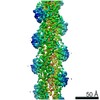 9734MC M: map data used to model this data C: citing same article ( |
|---|---|
| Similar structure data |
-Links
 PDBj
PDBj


- Assembly
| Deposited unit | 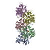
|
|---|---|
| 1 |
|
-Components
| #1: Protein | Mass: 41211.121 Da / Num. of mol.: 5 / Source method: isolated from a natural source / Source: (natural) Plasmid details: The cultivar of maize (Zea mays) plants is longping No.206. References: UniProt: B6TQ08 #2: Chemical | ChemComp-ADP /   Mass: 427.201 Da / Num. of mol.: 5 / Source method: obtained synthetically / Formula: C10H15N5O10P2 / Feature type: SUBJECT OF INVESTIGATION / Comment: ADP, energy-carrying molecule*YM Mass: 427.201 Da / Num. of mol.: 5 / Source method: obtained synthetically / Formula: C10H15N5O10P2 / Feature type: SUBJECT OF INVESTIGATION / Comment: ADP, energy-carrying molecule*YM#3: Chemical | ChemComp-MG /   Mass: 24.305 Da / Num. of mol.: 5 / Source method: obtained synthetically / Formula: Mg Mass: 24.305 Da / Num. of mol.: 5 / Source method: obtained synthetically / Formula: MgHas ligand of interest | Y | |
|---|
-Experimental details
-Experiment
| Experiment | Method: ELECTRON MICROSCOPY |
|---|---|
| EM experiment | Aggregation state: FILAMENT / 3D reconstruction method: helical reconstruction |
- Sample preparation
| Component | Name: The actin filament from Zea mays pollen / Type: COMPLEX Details: Plant actin was purified from the pollen which was collected from maize (Zea mays) plants and polymerized at room temperature for 4 hours. Entity ID: #1 / Source: NATURAL | ||||||||||||||||||||||||||||||||||||
|---|---|---|---|---|---|---|---|---|---|---|---|---|---|---|---|---|---|---|---|---|---|---|---|---|---|---|---|---|---|---|---|---|---|---|---|---|---|
| Molecular weight | Value: 15 kDa/nm | ||||||||||||||||||||||||||||||||||||
| Source (natural) | Organism: | ||||||||||||||||||||||||||||||||||||
| Buffer solution | pH: 7 | ||||||||||||||||||||||||||||||||||||
| Buffer component |
| ||||||||||||||||||||||||||||||||||||
| Specimen | Conc.: 0.4 mg/ml / Embedding applied: NO / Shadowing applied: NO / Staining applied: NO / Vitrification applied: YES Details: Plant actin was dialyzed against buffer solution with pH 7.0 overnight in order to change the alkaline pH to the neutral pH and then polymerized at the final concentration of 0.4 mg/mL. | ||||||||||||||||||||||||||||||||||||
| Specimen support | Grid material: COPPER / Grid mesh size: 400 divisions/in. | ||||||||||||||||||||||||||||||||||||
| Vitrification | Instrument: FEI VITROBOT MARK IV / Cryogen name: ETHANE / Humidity: 100 % / Chamber temperature: 298 K Details: Blotting time of 5.5 s and blotting force of level 2 |
- Electron microscopy imaging
| Experimental equipment |  Model: Titan Krios / Image courtesy: FEI Company |
|---|---|
| Microscopy | Model: FEI TITAN KRIOS |
| Electron gun | Electron source:  FIELD EMISSION GUN / Accelerating voltage: 300 kV / Illumination mode: FLOOD BEAM FIELD EMISSION GUN / Accelerating voltage: 300 kV / Illumination mode: FLOOD BEAM |
| Electron lens | Mode: BRIGHT FIELD / Nominal magnification: 22500 X / Calibrated magnification: 22013 X / Calibrated defocus min: 1200 nm / Calibrated defocus max: 2000 nm / Cs: 2.7 mm / C2 aperture diameter: 50 µm / Alignment procedure: COMA FREE |
| Specimen holder | Cryogen: NITROGEN / Specimen holder model: FEI TITAN KRIOS AUTOGRID HOLDER |
| Image recording | Average exposure time: 5.44 sec. / Electron dose: 39 e/Å2 / Detector mode: SUPER-RESOLUTION / Film or detector model: GATAN K2 SUMMIT (4k x 4k) / Num. of real images: 3605 Details: A total of 3,605 micrographs were recorded at a calibrated pixel size of 1.063 angstrom. |
| Image scans | Width: 7676 / Height: 7420 / Movie frames/image: 32 / Used frames/image: 2-27 |
- Processing
| Software | Name: PHENIX / Version: 1.11.1_2575: / Classification: refinement | |||||||||||||||||||||||||||||||||||||||||||||||||||||||
|---|---|---|---|---|---|---|---|---|---|---|---|---|---|---|---|---|---|---|---|---|---|---|---|---|---|---|---|---|---|---|---|---|---|---|---|---|---|---|---|---|---|---|---|---|---|---|---|---|---|---|---|---|---|---|---|---|
| EM software |
| |||||||||||||||||||||||||||||||||||||||||||||||||||||||
| Image processing | Details: The micrographs such as those with pollutions, bad Thon rings, large defocus values and others, were excluded before filament boxing. 1,540 micrographs were finally sorted out as "good" ones. | |||||||||||||||||||||||||||||||||||||||||||||||||||||||
| CTF correction | Type: PHASE FLIPPING AND AMPLITUDE CORRECTION | |||||||||||||||||||||||||||||||||||||||||||||||||||||||
| Helical symmerty | Angular rotation/subunit: -166.77 ° / Axial rise/subunit: 27.5 Å / Axial symmetry: C1 | |||||||||||||||||||||||||||||||||||||||||||||||||||||||
| Particle selection | Num. of particles selected: 40943 Details: A total of 8,609 ZMPA filaments were boxed using e2helixboxer.py in the package of EMAN2 with box width 168 and 77% box-overlap. A total of 40,943 segments were generated with box-size of 384. | |||||||||||||||||||||||||||||||||||||||||||||||||||||||
| 3D reconstruction | Resolution: 3.9 Å / Resolution method: FSC 0.143 CUT-OFF / Num. of particles: 35684 / Algorithm: BACK PROJECTION / Num. of class averages: 5100 / Symmetry type: HELICAL | |||||||||||||||||||||||||||||||||||||||||||||||||||||||
| Atomic model building | B value: 70 / Protocol: RIGID BODY FIT / Space: RECIPROCAL / Target criteria: Correlation coefficient Details: Chain C from pdb 5OOC was rigid body fitted to plant actin map using Chimera; Then Phenix was used for real space refinement. Finally, filament with 5 real space refinement units were ...Details: Chain C from pdb 5OOC was rigid body fitted to plant actin map using Chimera; Then Phenix was used for real space refinement. Finally, filament with 5 real space refinement units were generated using helical parameter, and this filament with 5 units was real space refined using Phenix again. | |||||||||||||||||||||||||||||||||||||||||||||||||||||||
| Atomic model building | PDB-ID: 5OOC Pdb chain-ID: C / Accession code: 5OOC / Source name: PDB / Type: experimental model | |||||||||||||||||||||||||||||||||||||||||||||||||||||||
| Refine LS restraints |
|