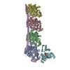
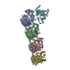
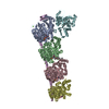
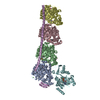
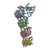
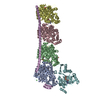
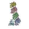
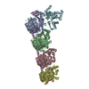
[English] 日本語
Yorodumi
- PDB-6y4n: Structure of Tubulin Tyrosine Ligase in Complex with Tb116 -

+
Open data


ID or keywords:

Loading...

-
Basic information

Entry
Database: PDB / ID: 6y4n
TitleStructure of Tubulin Tyrosine Ligase in Complex with Tb116
Components
  • Stathmin-4
  • Tubulin alpha-1B chain
  • Tubulin beta chain
  • Tubulin-Tyrosine Ligase
KeywordsLIGASE / TTL / Tubulin / complex
Function / homology
Function and homology information


tubulin-tyrosine ligase activity / Microtubule-dependent trafficking of connexons from Golgi to the plasma membrane / Hedgehog 'off' state / Cilium Assembly / Intraflagellar transport / COPI-dependent Golgi-to-ER retrograde traffic / Carboxyterminal post-translational modifications of tubulin / RHOH GTPase cycle / Sealing of the nuclear envelope (NE) by ESCRT-III / Kinesins ...tubulin-tyrosine ligase activity / Microtubule-dependent trafficking of connexons from Golgi to the plasma membrane / Hedgehog 'off' state / Cilium Assembly / Intraflagellar transport / COPI-dependent Golgi-to-ER retrograde traffic / Carboxyterminal post-translational modifications of tubulin / RHOH GTPase cycle / Sealing of the nuclear envelope (NE) by ESCRT-III / Kinesins / PKR-mediated signaling / The role of GTSE1 in G2/M progression after G2 checkpoint / Aggrephagy / Resolution of Sister Chromatid Cohesion / Mitotic Prometaphase / EML4 and NUDC in mitotic spindle formation / Separation of Sister Chromatids / RHO GTPases activate IQGAPs / RHO GTPases Activate Formins / Recruitment of NuMA to mitotic centrosomes / HSP90 chaperone cycle for steroid hormone receptors (SHR) in the presence of ligand / MHC class II antigen presentation / COPI-mediated anterograde transport / microtubule depolymerization / regulation of microtubule polymerization or depolymerization / tubulin binding / Hydrolases; Acting on acid anhydrides; Acting on GTP to facilitate cellular and subcellular movement / protein modification process / spindle microtubule / structural constituent of cytoskeleton / microtubule cytoskeleton organization / microtubule cytoskeleton / neuron projection development / mitotic cell cycle / growth cone / microtubule / neuron projection / nucleotide binding / GTPase activity / GTP binding / Golgi apparatus / metal ion binding / cytoplasm
Similarity search - Function
Tubulin-tyrosine ligase/Tubulin polyglutamylase / Tubulin-tyrosine ligase family / TTL domain profile. / Stathmin family / Stathmin, conserved site / Stathmin superfamily / Stathmin family / Stathmin family signature 1. / Stathmin family signature 2. / Stathmin-like (SLD) domain profile. ...Tubulin-tyrosine ligase/Tubulin polyglutamylase / Tubulin-tyrosine ligase family / TTL domain profile. / Stathmin family / Stathmin, conserved site / Stathmin superfamily / Stathmin family / Stathmin family signature 1. / Stathmin family signature 2. / Stathmin-like (SLD) domain profile. / Tubulin-beta mRNA autoregulation signal. / Alpha tubulin / Beta tubulin, autoregulation binding site / Beta tubulin / Tubulin / Tubulin, C-terminal / Tubulin C-terminal domain / Tubulin, conserved site / Tubulin subunits alpha, beta, and gamma signature. / Tubulin/FtsZ family, C-terminal domain / Tubulin/FtsZ-like, C-terminal domain / Tubulin/FtsZ, C-terminal / Tubulin/FtsZ, 2-layer sandwich domain / Tubulin/FtsZ family, GTPase domain / Tubulin/FtsZ family, GTPase domain / Tubulin/FtsZ, GTPase domain / Tubulin/FtsZ, GTPase domain superfamily
Similarity search - Domain/homology
PHOSPHOMETHYLPHOSPHONIC ACID ADENYLATE ESTER / GUANOSINE-5'-DIPHOSPHATE / GUANOSINE-5'-TRIPHOSPHATE / (2~{R})-1-methylpiperidine-2-carboxylic acid / Chem-O9K / Chem-O9N / benzyl hydrogen carbonate / DI(HYDROXYETHYL)ETHER / TRIETHYLENE GLYCOL / VALINE ...PHOSPHOMETHYLPHOSPHONIC ACID ADENYLATE ESTER / GUANOSINE-5'-DIPHOSPHATE / GUANOSINE-5'-TRIPHOSPHATE / (2~{R})-1-methylpiperidine-2-carboxylic acid / Chem-O9K / Chem-O9N / benzyl hydrogen carbonate / DI(HYDROXYETHYL)ETHER / TRIETHYLENE GLYCOL / VALINE / Tubulin tyrosine ligase / Tubulin beta chain / Stathmin-4 / Tubulin alpha-1B chain
Similarity search - Component
Biological speciesRattus norvegicus (Norway rat)
Gallus gallus (chicken)
Sus scrofa (pig)
MethodX-RAY DIFFRACTION / SYNCHROTRON / FOURIER SYNTHESIS / Resolution: 2.852 Å
AuthorsGavrilyuk, J. / Nocek, B. / Rigol, S. / Nicolaou, K.C. / Stoll, V.
CitationJournal: J.Org.Chem. / Year: 2021
Title: Design, Synthesis, and Biological Evaluation of Tubulysin Analogues, Linker-Drugs, and Antibody-Drug Conjugates, Insights into Structure-Activity Relationships, and Tubulysin-Tubulin Binding ...Title: Design, Synthesis, and Biological Evaluation of Tubulysin Analogues, Linker-Drugs, and Antibody-Drug Conjugates, Insights into Structure-Activity Relationships, and Tubulysin-Tubulin Binding Derived from X-ray Crystallographic Analysis.
Authors: Nicolaou, K.C. / Pan, S. / Pulukuri, K.K. / Ye, Q. / Rigol, S. / Erande, R.D. / Vourloumis, D. / Nocek, B.P. / Munneke, S. / Lyssikatos, J. / Valdiosera, A. / Gu, C. / Lin, B. / Sarvaiaya, H. ...Authors: Nicolaou, K.C. / Pan, S. / Pulukuri, K.K. / Ye, Q. / Rigol, S. / Erande, R.D. / Vourloumis, D. / Nocek, B.P. / Munneke, S. / Lyssikatos, J. / Valdiosera, A. / Gu, C. / Lin, B. / Sarvaiaya, H. / Trinidad, J. / Sandoval, J. / Lee, C. / Hammond, M. / Aujay, M. / Taylor, N. / Pysz, M. / Purcell, J.W. / Gavrilyuk, J.
History
DepositionFeb 21, 2020Deposition site: PDBE / Processing site: PDBE
Revision 1.0Mar 31, 2021Provider: repository / Type: Initial release
Revision 1.1Nov 23, 2022Group: Database references / Category: citation / citation_author / database_2
Item: _citation.country / _citation.journal_abbrev ..._citation.country / _citation.journal_abbrev / _citation.journal_id_ASTM / _citation.journal_id_CSD / _citation.journal_id_ISSN / _citation.journal_volume / _citation.page_first / _citation.page_last / _citation.pdbx_database_id_DOI / _citation.pdbx_database_id_PubMed / _citation.title / _citation.year / _database_2.pdbx_DOI / _database_2.pdbx_database_accession
Revision 1.2Jun 19, 2024Group: Data collection / Category: chem_comp_atom / chem_comp_bond

-
Structure visualization

Structure viewerMolecule:
MolmilJmol/JSmol

Downloads & links

-
Assembly

Deposited unit
A: Tubulin alpha-1B chain
B: Tubulin beta chain
C: Tubulin alpha-1B chain
D: Tubulin beta chain
E: Stathmin-4
F: Tubulin-Tyrosine Ligase
hetero molecules


Theoretical massNumber of molelcules
Total (without water)265,53726
Polymers261,4476
Non-polymers4,09020
Water10,827601
1


  • Idetical with deposited unit
  • defined by author&software
TypeNameSymmetry operationNumber
identity operation1_555x,y,z1
Buried area23690 Å2
ΔGint-113 kcal/mol
Surface area80660 Å2
MethodPISA
Unit cell
Length a, b, c (Å)104.284, 154.969, 183.433
Angle α, β, γ (deg.)90.000, 90.000, 90.000
Int Tables number19
Space group name H-MP212121

-
Components

-
Protein , 4 types, 6 molecules ACBDEF

#1: Protein Tubulin alpha-1B chain / Alpha-tubulin ubiquitous / Tubulin K-alpha-1 / Tubulin alpha-ubiquitous chain


Mass: 50204.445 Da / Num. of mol.: 2 / Source method: isolated from a natural source / Source: (natural) Sus scrofa (pig) / References: UniProt: Q2XVP4
#2: Protein Tubulin beta chain / Beta-tubulin


Mass: 49907.770 Da / Num. of mol.: 2 / Source method: isolated from a natural source / Source: (natural) Sus scrofa (pig) / References: UniProt: P02554
#3: Protein Stathmin-4 / Stathmin-like protein B3 / RB3


Mass: 16844.162 Da / Num. of mol.: 1
Source method: isolated from a genetically manipulated source
Source: (gene. exp.) Rattus norvegicus (Norway rat) / Gene: Stmn4 / Production host: Escherichia coli (E. coli) / References: UniProt: P63043
#4: Protein Tubulin-Tyrosine Ligase


Mass: 44378.496 Da / Num. of mol.: 1
Source method: isolated from a genetically manipulated source
Source: (gene. exp.) Gallus gallus (chicken) / Gene: TTL / Production host: Escherichia coli (E. coli) / References: UniProt: E1BQ43

-
Non-polymers , 14 types, 621 molecules

#5: Chemical ChemComp-GTP / GUANOSINE-5'-TRIPHOSPHATE


Mass: 523.180 Da / Num. of mol.: 3 / Source method: obtained synthetically / Formula: C10H16N5O14P3 / Comment: GTP, energy-carrying molecule*YM
#6: Chemical
ChemComp-MG / MAGNESIUM ION


Mass: 24.305 Da / Num. of mol.: 5 / Source method: obtained synthetically / Formula: Mg
#7: Chemical ChemComp-CA / CALCIUM ION


Mass: 40.078 Da / Num. of mol.: 2 / Source method: obtained synthetically / Formula: Ca
#8: Chemical ChemComp-GDP / GUANOSINE-5'-DIPHOSPHATE


Type: RNA linking / Mass: 443.201 Da / Num. of mol.: 1 / Source method: obtained synthetically / Formula: C10H15N5O11P2 / Comment: GDP, energy-carrying molecule*YM
#9: Chemical ChemComp-MES / 2-(N-MORPHOLINO)-ETHANESULFONIC ACID


Mass: 195.237 Da / Num. of mol.: 1 / Source method: obtained synthetically / Formula: C6H13NO4S / Comment: pH buffer*YM
#10: Chemical ChemComp-O9B / (2~{R})-1-methylpiperidine-2-carboxylic acid


Mass: 143.184 Da / Num. of mol.: 1 / Source method: obtained synthetically / Formula: C7H13NO2
#11: Chemical ChemComp-O9K / [(1~{R},3~{R})-1-(4-methanoyl-1,3-thiazol-2-yl)-4-methyl-3-(methylamino)pentyl] ethanoate


Mass: 284.375 Da / Num. of mol.: 1 / Source method: obtained synthetically / Formula: C13H20N2O3S
#12: Chemical ChemComp-O9N / methyl (2~{S},4~{S})-2,4-bis(azanyl)-5-phenyl-pentanoate


Mass: 222.284 Da / Num. of mol.: 1 / Source method: obtained synthetically / Formula: C12H18N2O2
#13: Chemical ChemComp-PGE / TRIETHYLENE GLYCOL


Mass: 150.173 Da / Num. of mol.: 1 / Source method: obtained synthetically / Formula: C6H14O4
#14: Chemical ChemComp-VAL / VALINE


Type: L-peptide linking / Mass: 117.146 Da / Num. of mol.: 1 / Source method: obtained synthetically / Formula: C5H11NO2
#15: Chemical ChemComp-P6S / benzyl hydrogen carbonate


Mass: 152.147 Da / Num. of mol.: 1 / Source method: obtained synthetically / Formula: C8H8O3
#16: Chemical ChemComp-PEG / DI(HYDROXYETHYL)ETHER


Mass: 106.120 Da / Num. of mol.: 1 / Source method: obtained synthetically / Formula: C4H10O3
#17: Chemical ChemComp-ACP / PHOSPHOMETHYLPHOSPHONIC ACID ADENYLATE ESTER / ADENOSINE-5'-[BETA, GAMMA-METHYLENE]TRIPHOSPHATE


Mass: 505.208 Da / Num. of mol.: 1 / Source method: obtained synthetically / Formula: C11H18N5O12P3 / Comment: AMP-PCP, energy-carrying molecule analogue*YM
#18: Water ChemComp-HOH / water


Mass: 18.015 Da / Num. of mol.: 601 / Source method: isolated from a natural source / Formula: H2O

-
Details

Has ligand of interestY

-
Experimental details

-
Experiment

ExperimentMethod: X-RAY DIFFRACTION / Number of used crystals: 1

-
Sample preparation

CrystalDensity Matthews: 2.83 Å3/Da / Density % sol: 56.61 %
Crystal growTemperature: 293 K / Method: vapor diffusion, sitting drop / pH: 6
Details: 30 mM calcium chloride, 30 mM magnesium chloride, 0.10 M MES pH 6.0, 2.0% w/v PEG 4000, 5.0% w/v glycerol

-
Data collection

DiffractionMean temperature: 100 K / Serial crystal experiment: N
Diffraction sourceSource: SYNCHROTRON / Site: Diamond / Beamline: I04 / Wavelength: 0.97949 Å
DetectorType: DECTRIS PILATUS3 6M / Detector: PIXEL / Date: Dec 6, 2018
RadiationProtocol: SINGLE WAVELENGTH / Monochromatic (M) / Laue (L): M / Scattering type: x-ray
Radiation wavelengthWavelength: 0.97949 Å / Relative weight: 1
ReflectionResolution: 2.852→78.93 Å / Num. obs: 69959 / % possible obs: 100 % / Redundancy: 6.7 % / Biso Wilson estimate: 79.07 Å2 / Rmerge(I) obs: 0.131 / Rsym value: 0.131 / Net I/σ(I): 11.1
Reflection shellResolution: 2.852→2.901 Å / Redundancy: 6.4 % / Rmerge(I) obs: 0.864 / Mean I/σ(I) obs: 2.1 / Num. unique obs: 3461 / Rsym value: 0.864 / % possible all: 100

-
Processing

Software
NameVersionClassification
BUSTER2.11.7 (6-FEB-2020)refinement
XDSdata reduction
Aimlessdata scaling
PDB_EXTRACT3.25data extraction
RefinementMethod to determine structure: FOURIER SYNTHESIS / Resolution: 2.852→78.929 Å / Cor.coef. Fo:Fc: 0.935 / Cor.coef. Fo:Fc free: 0.91 / Cross valid method: THROUGHOUT / σ(F): 0 / SU Rfree Blow DPI: 0.318
RfactorNum. reflection% reflectionSelection details
Rfree0.22 3527 5.04 %RANDOM
Rwork0.1827 ---
obs0.1846 69959 100 %-
Displacement parametersBiso max: 189.36 Å2 / Biso mean: 70.97 Å2 / Biso min: 25.06 Å2
Baniso -1Baniso -2Baniso -3
1--8.981 Å20 Å20 Å2
2---0.7938 Å20 Å2
3---9.7748 Å2
Refine analyzeLuzzati coordinate error obs: 0.33 Å
Refinement stepCycle: final / Resolution: 2.852→78.929 Å
ProteinNucleic acidLigandSolventTotal
Num. atoms17464 0 264 589 18317
Biso mean--60.29 53.08 -
Num. residues----2207
Refine LS restraints
Refine-IDTypeNumberRestraint functionWeightDev ideal
X-RAY DIFFRACTIONt_dihedral_angle_d10628SINUSOIDAL2
X-RAY DIFFRACTIONt_trig_c_planes
X-RAY DIFFRACTIONt_gen_planes5788HARMONIC5
X-RAY DIFFRACTIONt_it18202HARMONIC10
X-RAY DIFFRACTIONt_nbd
X-RAY DIFFRACTIONt_improper_torsion
X-RAY DIFFRACTIONt_pseud_angle
X-RAY DIFFRACTIONt_chiral_improper_torsion2333SEMIHARMONIC5
X-RAY DIFFRACTIONt_sum_occupancies
X-RAY DIFFRACTIONt_utility_distance
X-RAY DIFFRACTIONt_utility_angle
X-RAY DIFFRACTIONt_utility_torsion
X-RAY DIFFRACTIONt_ideal_dist_contact25894SEMIHARMONIC4
X-RAY DIFFRACTIONt_bond_d35299HARMONIC20.008
X-RAY DIFFRACTIONt_angle_deg63524HARMONIC20.97
X-RAY DIFFRACTIONt_omega_torsion3.07
X-RAY DIFFRACTIONt_other_torsion15.59
LS refinement shellResolution: 2.852→2.87 Å / Rfactor Rfree error: 0
RfactorNum. reflection% reflection
Rfree0.2587 69 4.93 %
Rwork0.241 1331 -
obs--100 %
Refinement TLS params.

Method: refined / Refine-ID: X-RAY DIFFRACTION

IDL112)L122)L132)L222)L232)L332)S11 (Å °)S12 (Å °)S13 (Å °)S21 (Å °)S22 (Å °)S23 (Å °)S31 (Å °)S32 (Å °)S33 (Å °)T112)T122)T132)T222)T232)T332)Origin x (Å)Origin y (Å)Origin z (Å)
10.05920.3338-0.01131.309-0.92131.3493-0.01980.00640.078-0.05280.1589-0.05970.2057-0.1705-0.1391-0.0133-0.0177-0.039-0.04340.0954-0.0508-25.3161-4.320261.951
20.2304-0.1615-0.02960.7934-0.77291.1635-0.0024-0.0297-0.014-0.0868-0.0105-0.02840.09250.02620.0129-0.05390.0063-0.0012-0.01820.0520.0003-15.467620.913329.6166
30.1744-0.0099-0.21680.4963-0.40810.5447-0.10450.0211-0.0377-0.03390.0518-0.0248-0.03630.00590.0527-0.0336-0.05030.0074-0.00150.0190-12.589749.1815-3.7711
40.74480.03330.18680.8328-0.51580.7777-0.27940.31510.0647-0.01560.17350.035-0.15980.19270.10590.0103-0.152-0.08820.07330.152-0.1434-16.936580.4007-31.6036
50.1371-0.27680.4752.3624-2.91043.6232-0.0814-0.0215-0.00890.19780.38390.33-0.2348-0.5442-0.3025-0.08560.0347-0.02870.05680.152-0.0166-39.244538.028120.0263
61.0095-0.07661.15160.06510.43212.5932-0.3075-0.06620.27590.00580.1034-0.0919-0.2539-0.24630.2040.13910.0813-0.152-0.1797-0.0618-0.0129-5.521524.065292.5146
Refinement TLS group
IDRefine-IDRefine TLS-IDSelection detailsAuth asym-IDAuth seq-ID
1X-RAY DIFFRACTION1{ A|* }A1 - 439
2X-RAY DIFFRACTION2{ B|* }B1 - 431
3X-RAY DIFFRACTION3{ C|* }C1 - 440
4X-RAY DIFFRACTION4{ D|* }D1 - 431
5X-RAY DIFFRACTION5{ E|* }E50 - 187
6X-RAY DIFFRACTION6{ F|* }F1 - 381

+
About Yorodumi

-
News

-
Feb 9, 2022. New format data for meta-information of EMDB entries

New format data for meta-information of EMDB entries

  • Version 3 of the EMDB header file is now the official format.
  • The previous official version 1.9 will be removed from the archive.

Related info.:EMDB header

External links:wwPDB to switch to version 3 of the EMDB data model

-
Aug 12, 2020. Covid-19 info

Covid-19 info

URL: https://pdbjlvh1.pdbj.org/emnavi/covid19.php

New page: Covid-19 featured information page in EM Navigator.

Related info.:Covid-19 info / Mar 5, 2020. Novel coronavirus structure data

+
Mar 5, 2020. Novel coronavirus structure data

Novel coronavirus structure data

Related info.:Yorodumi Speices / Aug 12, 2020. Covid-19 info

External links:COVID-19 featured content - PDBj / Molecule of the Month (242):Coronavirus Proteases

+
Jan 31, 2019. EMDB accession codes are about to change! (news from PDBe EMDB page)

EMDB accession codes are about to change! (news from PDBe EMDB page)

  • The allocation of 4 digits for EMDB accession codes will soon come to an end. Whilst these codes will remain in use, new EMDB accession codes will include an additional digit and will expand incrementally as the available range of codes is exhausted. The current 4-digit format prefixed with “EMD-” (i.e. EMD-XXXX) will advance to a 5-digit format (i.e. EMD-XXXXX), and so on. It is currently estimated that the 4-digit codes will be depleted around Spring 2019, at which point the 5-digit format will come into force.
  • The EM Navigator/Yorodumi systems omit the EMD- prefix.

Related info.:Q: What is EMD? / ID/Accession-code notation in Yorodumi/EM Navigator

External links:EMDB Accession Codes are Changing Soon! / Contact to PDBj

+
Jul 12, 2017. Major update of PDB

Major update of PDB

  • wwPDB released updated PDB data conforming to the new PDBx/mmCIF dictionary.
  • This is a major update changing the version number from 4 to 5, and with Remediation, in which all the entries are updated.
  • In this update, many items about electron microscopy experimental information are reorganized (e.g. em_software).
  • Now, EM Navigator and Yorodumi are based on the updated data.

External links:wwPDB Remediation / Enriched Model Files Conforming to OneDep Data Standards Now Available in the PDB FTP Archive

-
Yorodumi

Thousand views of thousand structures

  • Yorodumi is a browser for structure data from EMDB, PDB, SASBDB, etc.
  • This page is also the successor to EM Navigator detail page, and also detail information page/front-end page for Omokage search.
  • The word "yorodu" (or yorozu) is an old Japanese word meaning "ten thousand". "mi" (miru) is to see.

Related info.:EMDB / PDB / SASBDB / Comparison of 3 databanks / Yorodumi Search / Aug 31, 2016. New EM Navigator & Yorodumi / Yorodumi Papers / Jmol/JSmol / Function and homology information / Changes in new EM Navigator and Yorodumi

Read more